Brux Checker® Foil: A Great Way to See Tooth Wear Patterns

June 26, 2023 Lee Ann Brady DMD

Sometimes we suspect a dental patient has tooth wear or damage from attrition. In these cases, we want to help the patient understand what they do with their teeth while sleeping. We also want to see for ourselves the patterns of wear. I recently learned about the Brux Checker® diagnostic material during the 2022 Masters Week at The Pankey Institute.

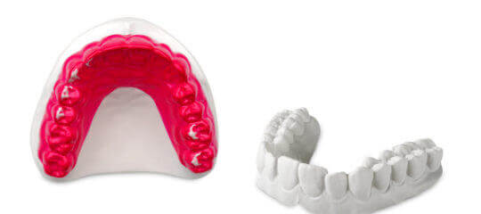
During Masters Week, Dr. Ricardo Armanetto from Genoa, Italy, showed us this material, which is 0.1 mm in thickness. One side of the material is red, and the other is foil. The material can be placed in your MiniStar®, BioStar®, or Vaquform thermoformer to create a suck-down device that your patient wears over their upper arch overnight. A suggestion is to make two of these devices and ask your patient to wear one for one night and one for a second night.

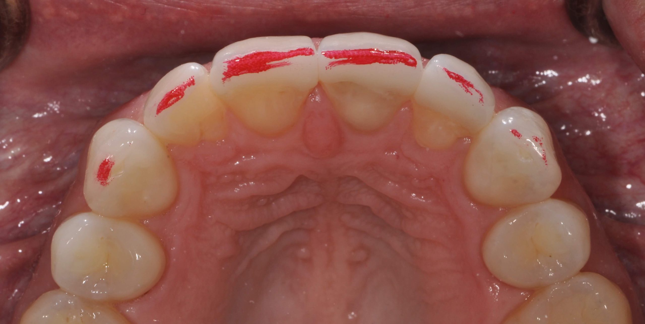
If the patient is para-functioning during sleep, they will wear the red off the device in the places where their teeth are touching within 0.1 mm of each other. You will see which teeth are touching and grinding.

In thinking about using Brux Checker, the following cases came to mind:

  • Brux Checker is designed as a patient education tool. I want to use Brux Checker for patients I think are para-functioning because of signs of wear and attrition—and now I need them to take some ownership of their parafunction. This is an easy, inexpensive way to do that, in addition to the QuickSplints I use in my practice.
  • I also want to use Brux Checker with patients I have equilibrated to double-check my equilibration. Sometimes, when the patient is in the dental chair, it is difficult for the patient to find a posterior interference that I failed to clear out. I want to ensure they are not damaging their teeth while para functioning on molars during sleep.
  • Similarly, after placing dental restorations, I can use Brux Checker to fine-tune the occlusion after seeing what happens during the night versus in my dental chair.
  • In the case of Class IV corners or incisal edge repairs that people want to be replaced in composite and the composite pops off, they may not know that they parafunction and there is a need to fine-tune their occlusion. I don’t know if I can get one of these patients to wear a Brux Checker during the daytime, but it should be easy to get them to wear one of these devices during the night. If the red wears off the foil at the spot where the composite has been used to repair a Class IV corner or incisal edge, there will be no question about the stress they are placing on the repair.

You can see that I’m thinking beyond patient education to using Brux Checker to help me fine-tune someone’s occlusion. I know there are places where people bring their teeth together at night that they can’t show me in the chair.

Related Course

E1: Aesthetic & Functional Treatment Planning

DATE: March 13 2025 @ 8:00 am - March 16 2025 @ 2:30 pm

Location: The Pankey Institute

CE HOURS: 39

Dentist Tuition: $ 6800

Single Occupancy with Ensuite Private Bath (Per Night): $ 345

Transform your experience of practicing dentistry, increase predictability, profitability and fulfillment. The Essentials Series is the Key, and Aesthetic and Functional Treatment Planning is where your journey begins.  Following a system of…

Learn More>

About Author

User Image
Lee Ann Brady DMD

Dr. Lee Ann Brady is passionate about dentistry, her family and making a difference. She is a general dentist and owns a practice in Glendale, AZ limited to restorative dentistry. Lee’s passion for dental education began as a CE junkie herself, pursuing lots of advanced continuing education focused on Restorative and Occlusion. In 2005, she became a full time resident faculty member for The Pankey Institute, and was promoted to Clinical Director in 2006. Lee joined Spear Education as Executive VP of Education in the fall of 2008 to teach and coordinate the educational curriculum. In June of 2011, she left Spear Education, founded leeannbrady.com and joined the dental practice she now owns as an associate. Today, she teaches at dental meetings and study clubs both nationally and internationally, continues to write for dental journals and her website, sits on the editorial board of the Journal of Cosmetic Dentistry, Inside Dentistry and DentalTown Magazines and is the Director of Education for The Pankey Institute.

FIND A PANKEY DENTIST OR TECHNICIAN

I AM A
I AM INTERESTED IN

VIEW COURSE CALENDAR

Incisal Edge Anatomy

November 30, 2017 Lee Ann Brady DMD

Anterior teeth have a complex incisal edge anatomy that creates both the esthetic appearance of the tooth and the function of the upper and lower incisors against one another.

Often in both ceramics and in composite we do not recreate the full anatomic form of the tooth. This results in both esthetic and functional challenges for the patient.  When we look at incisal edges from a lateral perspective there are three components, the pitch and two bevels. The Pitch is the flat top of the incisal edge. On both the labial and the lingual the transition zone between the pitch and these surfaces is a bevel. One is referred to as the leading edge and one is referred to as the trailing edge.

The Pitch has dimension or labio-lingual width, usually at least 1mm. This width increases as the patient shortens the tooth from Attrition, if they parafunction in an edge to edge position. The pitch is not always parallel to the horizon, but it’s relative position depends on the inclination of the incisor. When the incisors are optimally inclined, just slightly further to the labial at the incisal edge the pitch is slanted upward toward the lingual. This creates the incisal edge esthetic effect of thinner enamel at the labio-incisal junction and creates visual translucence. If the pitch is level to the horizon it changes light reflection and the appearance of the tooth. These two factors together is often what changes in restorative material.

We create a pitch that is level to the horizon, and then to gain translucence we decrease the width of the pitch, sometimes to a knife edge.

The challenge with this, is that patients sit in edge to edge position often to incise food and some for parafunction. If there is insufficient width to the pitch they may experience functional challenges.

The bevels on both sides have a variable width, but can be between a portion of a millimeter to multiple millimeters long. The bevels get longer in patients who grind their teeth in an excursive pathway pattern. Patients who parafunction edge to edge can often eliminate the bevel, making it easier to shear off enamel on the labial or lingual side of the tooth, or chip the edge enamel. The bevel functionally is a transition zone to create smooth functional movement as we pass from excursive movements onto the pitch. The Intercuspal stops on lower incisors is often on or gingival to the bevel.

Whether we are finalizing an equilibration or finalizing the occlusion on composites or ceramics perfecting the anterior guidance requires both pitch and bevel surfaces, it is a perfect example of the marriage between form and function.

Related Course

Utilizing Clear Aligner Therapy in a Digital Workflow

DATE: September 26 2024 @ 6:00 pm - September 28 2024 @ 2:00 pm

Location: The Pankey Institute

CE HOURS: 0

Dentist Tuition: $ 1695

Single Occupancy with Ensuite Private Bath (per night): $ 290

Enhance Restorative Outcomes This one-and-a-half-day course is designed for Invisalign providers who want to improve restorative outcomes with clear aligner therapy. The course focuses on the digital workflow for comprehensive…

Learn More>

About Author

User Image
Lee Ann Brady DMD

Dr. Lee Ann Brady is passionate about dentistry, her family and making a difference. She is a general dentist and owns a practice in Glendale, AZ limited to restorative dentistry. Lee’s passion for dental education began as a CE junkie herself, pursuing lots of advanced continuing education focused on Restorative and Occlusion. In 2005, she became a full time resident faculty member for The Pankey Institute, and was promoted to Clinical Director in 2006. Lee joined Spear Education as Executive VP of Education in the fall of 2008 to teach and coordinate the educational curriculum. In June of 2011, she left Spear Education, founded leeannbrady.com and joined the dental practice she now owns as an associate. Today, she teaches at dental meetings and study clubs both nationally and internationally, continues to write for dental journals and her website, sits on the editorial board of the Journal of Cosmetic Dentistry, Inside Dentistry and DentalTown Magazines and is the Director of Education for The Pankey Institute.

FIND A PANKEY DENTIST OR TECHNICIAN

I AM A
I AM INTERESTED IN

VIEW COURSE CALENDAR

Adult Growth of the Dental Arch

September 4, 2017 Roger Solow DDS

Successful restorative dentistry now hinges on an understanding that physiological changes occur over a lifetime. It’s detrimental to treat the dynamic relationship between dental occlusion and adjacent craniofacial structures as static.

We are all generally familiar with the fact that there is a significant change in facial profile (convex to straight) between adolescence and adulthood. Jaw growth usually ends between 17 and 20-ish years old, but 3-dimensional craniofacial skeletal growth and remodeling does not cease after adolescence.

It’s lifelong growth even though it’s slow. As a result, we can’t consider adult patients morphologically stable. This is actually a relatively new concept that we’ve become aware of because of implant dentistry.

So what does this mean for restorations? First, we need more information.

Physiological Changes and Restorative Dentistry: A Quick Overview

These adult growth changes can be seen in both a decrease and increase in the dimensions of the craniofacial skeleton. There is an increase in maxillary and mandibular anterior dentoalveolar heights.

We should also pay attention to vertical growth of the maxilla, which continues after transverse and sagittal growth end. It has been suggested that reductions seen in arch width, depth, and perimeter may be due to interstitial wear and mesial drift. The latter occurs because of an occlusal force stemming from root angulation, mesial eruption force and the direction of occlusal contact during chewing. It’s integral to consider tooth movement because it compensates for wear while maintaining interproximal contacts.

There are different patterns of growth in short-faced and long-faced people. Short-faced individuals have greater transverse maxillary growth. As they mature, their anterior teeth tip forward and enable mesial drift. This process occurs more vertically in long-faced people. Short-faced individuals experience upward buccal movement of the teeth, while long-faced individuals experience lingual movement and continual tooth eruption that supports a normal interarch relationship.

What we now know from recent research is that eruption after the tooth has reached occlusal contact is a compensatory response to occlusal wear. Eruption creates vertical growth if there is no occlusal wear.

A comprehensive understanding of the complex interplay between all of these changes in the dental arch is essential to restorative dentistry.

How do you keep up to date on the latest dental research? We’d love to hear your tips in the comments! 

Related Course

Masters’ Week

DATE: June 2 2025 @ 8:00 am - June 5 2025 @ 2:00 pm

Location: The Pankey Institute

CE HOURS: 27

Dentist Tuition: $ 8500

Single Occupancy with Ensuite Private Bath (per night): $ 345

Master Your Skills Masters’ Week is a unique learning experience each and every year where we bring together a group of talented speakers to share on a range of topics….

Learn More>

About Author

User Image
Roger Solow DDS

Roger Solow received a BA in Biology from UCLA in 1975 and his DDS with honors from University of the Pacific School of Dentistry in 1978. He is a general dentist and has a full time, fee-for-service practice that he limits to restorative dentistry in Mill Valley, California. He is a Pankey Scholar and a lead visiting faculty at the Pankey Institute in Key Biscayne, Florida. He has taught restorative dentistry at UOP Dental School and has lectured to study clubs, dental societies, and the national meetings of the Academy of General Dentistry. Dr. Solow is a Fellow of the American College of Dentistry. Dr. Solow is a frequently published author.

FIND A PANKEY DENTIST OR TECHNICIAN

I AM A
I AM INTERESTED IN

VIEW COURSE CALENDAR