When Your Occlusal Clearance Disappears

April 19, 2019 Lee Ann Brady DMD

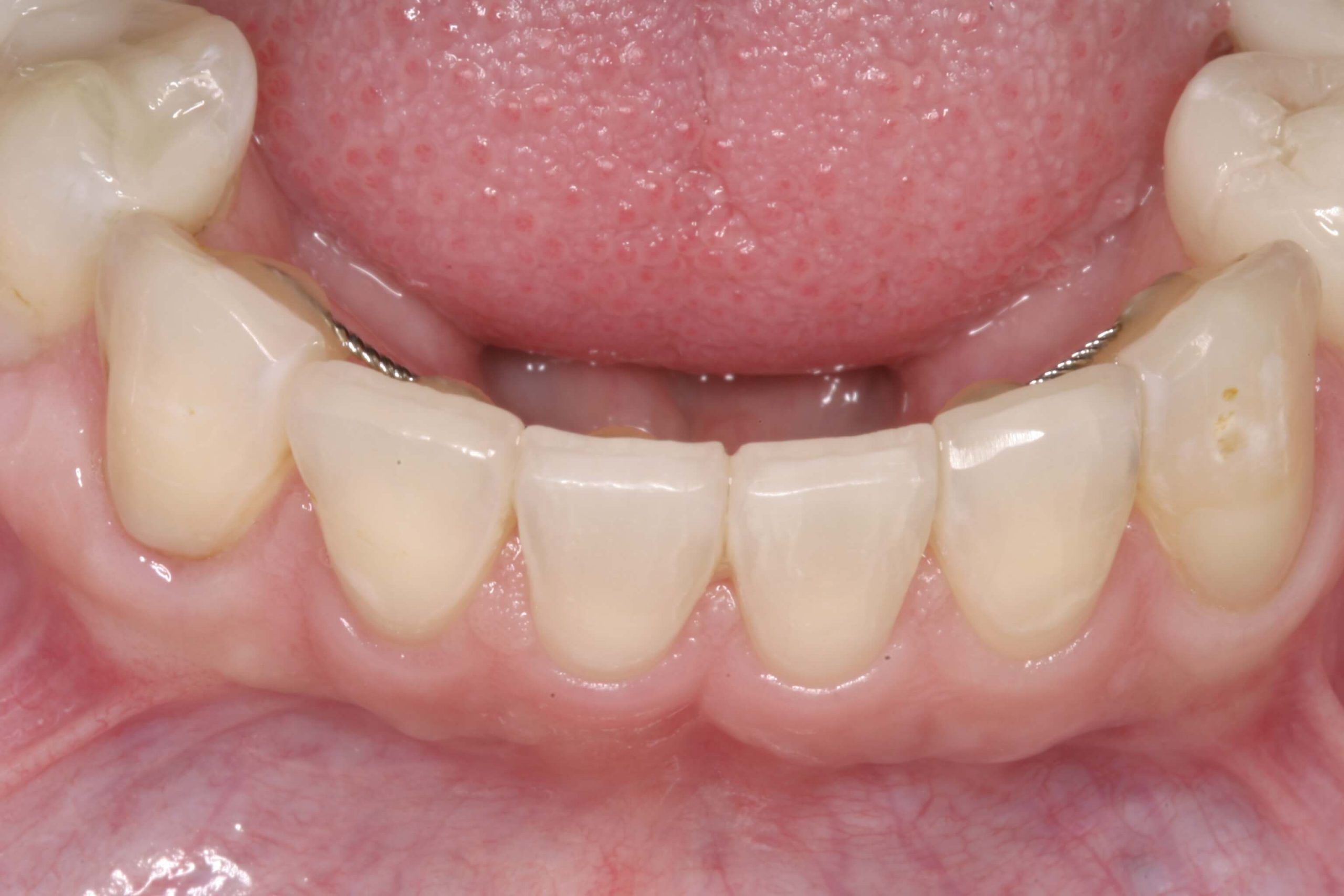
It can be an incredibly frustrating clinical situation, when you have been meticulous about preparing a posterior tooth, (most commonly a molar) for a crown and things aren’t predictable. Using your burs you created depth cuts to ensure adequate occlusal clearance. After the impression you allow your assistant to fabricate the temporary only to have them come get you. Why? Because the temp is thin or perforated on the occlusal. When you go back to check, and have the patient bite, sure enough the opposing tooth is touching your prep.

A common reason that this happens is because we just prepared away the patient’s first point of contact in centric relation. The lateral pterygoid muscle in coordination with the elevator muscles has a learned pattern of firing that bring the mandibular teeth into maximum intercuspal position. This “learned” position is programmed by the patient’s first point of contact when the condyles are seated. For some patients when we remove this contact, and therefore the message that was programming the muscles to locate MIP, they release quickly. When the muscles release and the condyles seat, the occlusion is now totally different than MIP was moments before.

Leaf Gauge
Finding First Point of Contact

One solution that I considered briefly was to no longer work on molars! Alas, not a great business strategy.

Removing this frustration is about understanding which patients are at risk. Identifying risk begins with the exam, whether we are discussing caries or occlusion. There are several key factors that alert me to this potential issue. I start by identifying the patient’s first point of contact and clarifying if it is on the tooth we are about to prepare. If I am going to prepare FPC away, then I look at the magnitude and direction of the patient’s slide, or the difference between this position and MIP. If the difference is small (1-2mm), then even if their condyle does seat the occlusal difference will not cause an issue for clearance. So large slides (3mm or greater) could cost approximately 1mm of clearance on the prepared tooth. Other factors include whether they have a history of occlusal changes or more than one MIP they can find.

Understanding the risk, still leaves us with the question of how to proceed. That is a longer conversation for another post. However, if we proceed as we would before, at least knowing the risk we can explain this to the patient ahead of time, and help them understand how we would manage it if it happens.

Related Course

E1: Aesthetic & Functional Treatment Planning

DATE: January 23 2025 @ 8:00 am - January 26 2025 @ 2:30 pm

Location: The Pankey Institute

CE HOURS: 39

Dentist Tuition: $ 6800

Single Occupancy with Ensuite Private Bath (Per Night): $ 345

Transform your experience of practicing dentistry, increase predictability, profitability and fulfillment. The Essentials Series is the Key, and Aesthetic and Functional Treatment Planning is where your journey begins.  Following a system of…

Learn More>

About Author

User Image
Lee Ann Brady DMD

Dr. Lee Ann Brady is passionate about dentistry, her family and making a difference. She is a general dentist and owns a practice in Glendale, AZ limited to restorative dentistry. Lee’s passion for dental education began as a CE junkie herself, pursuing lots of advanced continuing education focused on Restorative and Occlusion. In 2005, she became a full time resident faculty member for The Pankey Institute, and was promoted to Clinical Director in 2006. Lee joined Spear Education as Executive VP of Education in the fall of 2008 to teach and coordinate the educational curriculum. In June of 2011, she left Spear Education, founded leeannbrady.com and joined the dental practice she now owns as an associate. Today, she teaches at dental meetings and study clubs both nationally and internationally, continues to write for dental journals and her website, sits on the editorial board of the Journal of Cosmetic Dentistry, Inside Dentistry and DentalTown Magazines and is the Director of Education for The Pankey Institute.

FIND A PANKEY DENTIST OR TECHNICIAN

I AM A
I AM INTERESTED IN

VIEW COURSE CALENDAR

Esthetics & Function: Incisal Edge Bevels

December 10, 2017 Lee Ann Brady DMD

There are three critical components to the incisal edge anatomy of anterior teeth. Understanding the function and esthetics of the pitch and two bevels is essential to creating an ideal patient result.

How can a clinician re-create the full anatomic form of the tooth in ceramics and composite? 

In my last blog on this topic, I discussed the dimensions, characterization, esthetics, restorative approach and challenge of mimicking ‘pitch‘ esthetics. Now, I’ll delve into mastering the bevels to create superior restorative results. Combining an esthetic pitch with functional bevels will ensure a smooth composite or ceramic outcome.

Components of Incisal Edge Anatomy Function and Esthetics: Bevels

The two bevels can be found on alternately the labial and the lingual of the transition zone between the pitch and these surfaces. They are often called the leading edge and the trailing edge.

Bevels

Dimensions: The bevels on both sides have a variable width. They can be between less than a millimeter to multiple millimeters long.

Characterization: The bevels lengthen in patients who grind their teeth in an excursive pathway pattern. Patients who parafunction edge to edge might eliminate the bevel. This makes it easier to shear enamel off on the labial or lingual side of the tooth. It also could result in chipping the edge enamel.

Function: The bevel is a transition zone to create smooth functional movement passing from excursive movements onto the pitch. Intercuspal stops on lower incisors are often on or gingival to the bevel.

Whether you are finalizing an equilibration, the occlusion on composites, or ceramics, perfecting anterior guidance is all about both pitch and bevel surfaces. These critical components are a great example of marrying form and function in your technique.

What is your restorative approach for recreating incisal edge anatomy? We’d love to hear from you in the comments! 

Related Course

E1: Aesthetic & Functional Treatment Planning

DATE: December 11 2025 @ 8:00 am - December 14 2025 @ 2:30 pm

Location: The Pankey Institute

CE HOURS: 39

Dentist Tuition: $ 6800

Single Occupancy with Ensuite Private Bath (Per Night): $ 345

Transform your experience of practicing dentistry, increase predictability, profitability and fulfillment. The Essentials Series is the Key, and Aesthetic and Functional Treatment Planning is where your journey begins.  Following a system of…

Learn More>

About Author

User Image
Lee Ann Brady DMD

Dr. Lee Ann Brady is passionate about dentistry, her family and making a difference. She is a general dentist and owns a practice in Glendale, AZ limited to restorative dentistry. Lee’s passion for dental education began as a CE junkie herself, pursuing lots of advanced continuing education focused on Restorative and Occlusion. In 2005, she became a full time resident faculty member for The Pankey Institute, and was promoted to Clinical Director in 2006. Lee joined Spear Education as Executive VP of Education in the fall of 2008 to teach and coordinate the educational curriculum. In June of 2011, she left Spear Education, founded leeannbrady.com and joined the dental practice she now owns as an associate. Today, she teaches at dental meetings and study clubs both nationally and internationally, continues to write for dental journals and her website, sits on the editorial board of the Journal of Cosmetic Dentistry, Inside Dentistry and DentalTown Magazines and is the Director of Education for The Pankey Institute.

FIND A PANKEY DENTIST OR TECHNICIAN

I AM A
I AM INTERESTED IN

VIEW COURSE CALENDAR