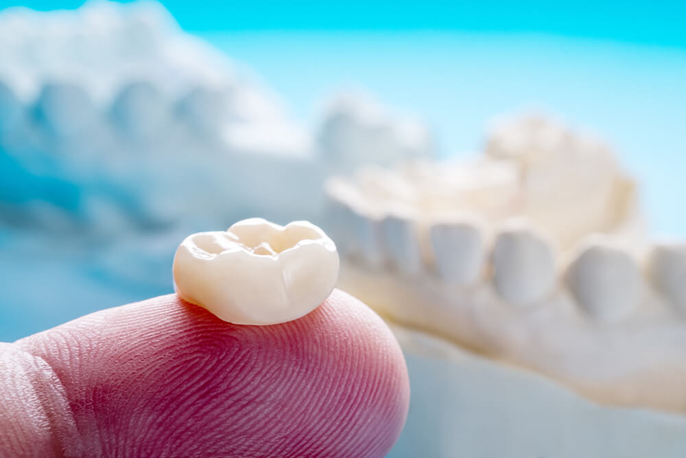
Not Every Endodontically Treated Tooth Needs a Crown

August 22, 2022 Lee Ann Brady DMD

Not every tooth that has been endodontically treated requires a crown to insure it has great longevity and doesn’t crack or fracture.

Molars

Very clearly the literature supports that molar teeth in the posterior absolutely must have four cusp coverage—a four-cusp onlay or a full coverage crown. We are trying to use the phenomenon of containment with strong ceramic or metal material around the entire circumference of the tooth. We are holding the buccal and lingual together and replacing the top of the root chamber, so the tooth doesn’t fracture.

Bicuspids

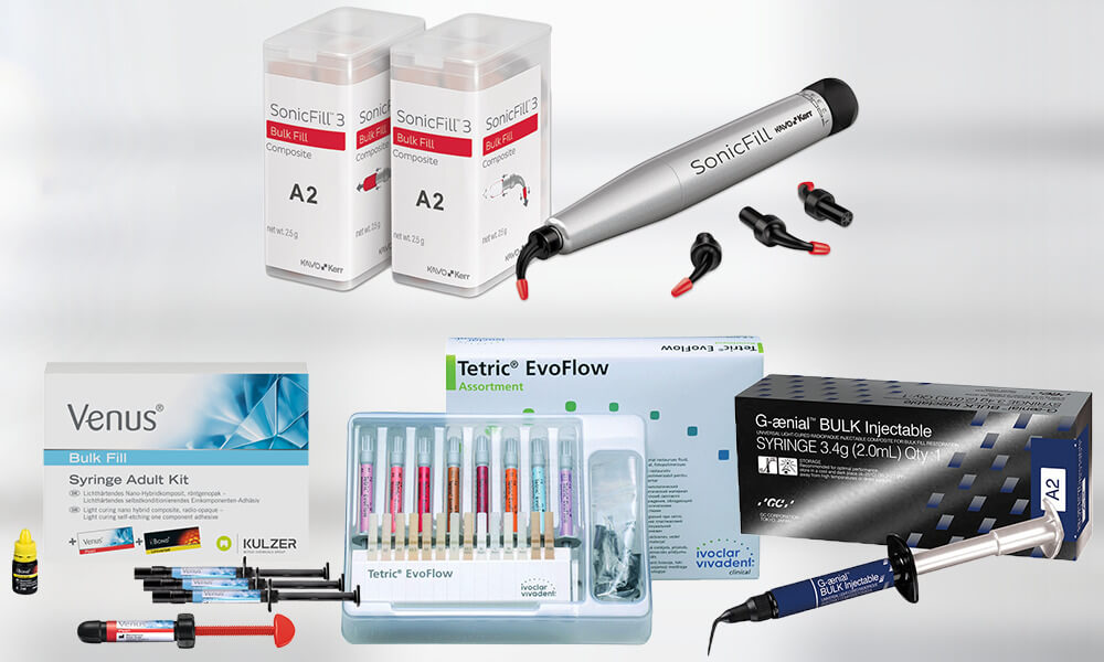
Bicuspids that have been endodontically treated do not need two-cusp coverage if there have been no previous restorations and the endo access is very conservative. In the case of a premolar that has never had an MO, a DO, or an MOD, and has a tiny access hole, you can do a composite buildup or chamber retaining composite restoration. If the patient has high functional risk, a reasonable decision would be to restore the tooth with an onlay or crown

Anterior Teeth

There is no scientific support for doing a crown on an anterior tooth just because it has had endodontic therapy. We do a crown on an anterior tooth that has had endo when it is already structurally compromised, for example with previous mesial lingual and distal lingual composite fillings, missing tooth structure, and significant structural compromise between the endo access and other restorations.

Related Course

E1: Aesthetic & Functional Treatment Planning

DATE: June 20 2024 @ 8:00 am - June 23 2024 @ 2:30 pm

Location: The Pankey Institute

CE HOURS: 39

Dentist Tuition: $ 6500

Single Occupancy Room with Ensuite Bath (Per Night): $ 290

Transform your experience of practicing dentistry, increase predictability, profitability and fulfillment. The Essentials Series is the Key, and Aesthetic and Functional Treatment Planning is where your journey begins.  Following a system of…

Learn More>

About Author

User Image
Lee Ann Brady DMD

Dr. Lee Ann Brady is passionate about dentistry, her family and making a difference. She is a general dentist and owns a practice in Glendale, AZ limited to restorative dentistry. Lee’s passion for dental education began as a CE junkie herself, pursuing lots of advanced continuing education focused on Restorative and Occlusion. In 2005, she became a full time resident faculty member for The Pankey Institute, and was promoted to Clinical Director in 2006. Lee joined Spear Education as Executive VP of Education in the fall of 2008 to teach and coordinate the educational curriculum. In June of 2011, she left Spear Education, founded leeannbrady.com and joined the dental practice she now owns as an associate. Today, she teaches at dental meetings and study clubs both nationally and internationally, continues to write for dental journals and her website, sits on the editorial board of the Journal of Cosmetic Dentistry, Inside Dentistry and DentalTown Magazines and is the Director of Education for The Pankey Institute.

FIND A PANKEY DENTIST OR TECHNICIAN

I AM A
I AM INTERESTED IN

VIEW COURSE CALENDAR

Functional Risk Part 3 – Occlusal Therapy 

July 1, 2022 Lee Ann Brady DMD

Why Occlusal Appliance Therapy Is My First Step Prior to Ortho, Equilibration, or Restorative

Occlusal changes on an appliance are easy and reversible. An appliance can immediately reduce elevator muscle activity and give the patient relief. The patient also experiences what changes to their tooth contacts could provide for them long term. We can test the changes that would be made by ortho, equilibration, and/or restorative.

As reviewed in Part 2 of this series, our goals are to stabilize the joint anatomy and reduce the activity of the elevator muscles because those muscles are what overload the joints and teeth. We also want to slow down the rate of damage to the dentition and move that rate back to a more age-appropriate pace. We also may need to reorganize a patient’s occlusion to manage occlusal forces to ensure restorations that last.

Removing Posterior Contacts Does Not Work for Every Patient

Over my years of clinical practice, I have found that changing the occlusion does reduce functional risk for most patients. But we all have patients with perfect occlusion who present with TMD symptoms. We have some patients who continue to parafunction after we move them into immediate posterior disclusion.

Studies show that proprioception causes the elevator muscles to engage in only 80 to 85% of the population. This means that when the brain receives the signal that teeth are touching, the brain elevates the masseter muscles in 80 to 85% of people. Tooth contact is the trigger. Because this proprioception does not occur for 15 to 20% of the population, it is not the universal trigger for excessive loading.

Over my years in clinical practice, I have learned there is nothing I can do that is 100% dependable to stop a patient from para-functioning. Some of my patients continue to excessively load after posterior contacts are removed. Their functional risk does not diminish.

If we cannot reduce elevator force and redistribute force enough on an occlusal appliance to eliminate or at least relieve TMD symptoms, then occlusal therapy via ortho, equilibration, or restorative will not satisfactorily help the patient. We will need to turn to other forms of therapy.

Other modalities I use are BOTOX to deactivate muscles, massage therapy, and physical therapy. There are also systemic medications, cold lasers, and TENS therapy we can use to reduce the activity of the muscles or reduce inflammation in the muscles and joints. Sometimes one modality will alleviate symptoms for a while and when symptoms return, we can try it again or try another modality.

An Exercise to Identify the Patients Who May Not Benefit from Occlusal Therapy

You can do what I call a poor man’s EMG on yourself by placing your hands on your masseter muscles. Put your back teeth together, clench and release, clench and release, clench and release to see how much masseter activity you have. Then move your teeth into protrusive edge to edge and try to clench a little bit, making sure your back teeth do not touch. If you now have a posterior tooth touching in the edge-to-edge position, then put a pencil or pen between your front teeth to separate your back teeth.

With no back teeth touching and contact on the centrals, try to clench and release two or three times while feeling your masseters. Most of you will find your masseters do not move or move a lot less when no back teeth are touching. Some of you, even with your back teeth separated, can still clench in protrusive and can still increase the muscle activity almost the same amount as when your back teeth touch.

I do this exercise with my patients, but when they move into protrusive, I put a bite stop over their front teeth or have them bite on a Lucia jig we have lined for their bite registration. If you do this test with your patients, you can use an EMG or feel the muscle activity with your hands.

If the patient can still generate almost the same force or the same force with their back teeth separated, you have identified one of the around 15% of people who might not benefit significantly from occlusal therapy. You’ve also identified someone who might not do well on an anterior-only appliance because, if they can generate that same force on just two teeth, they are at risk for those teeth becoming sore and moving.

Interested in Learning More?

The Pankey Institute Essentials courses and multiple focus courses include hands-on exercises and over-the-shoulder training designed to help dentists develop mastery in reducing functional risk and treating TMD symptoms.

Related Course

E2: Occlusal Appliances & Equilibration

DATE: April 6 2025 @ 8:00 am - April 10 2025 @ 2:30 pm

Location: The Pankey Institute

CE HOURS: 44

Dentist Tuition: $ 7400

Single Occupancy with Ensuite Private Bath (per night): $ 345

What if you had one tool that increased comprehensive case acceptance, managed patients with moderate to high functional risk, verified centric relation and treated signs and symptoms of TMD? Appliance…

Learn More>

About Author

User Image
Lee Ann Brady DMD

Dr. Lee Ann Brady is passionate about dentistry, her family and making a difference. She is a general dentist and owns a practice in Glendale, AZ limited to restorative dentistry. Lee’s passion for dental education began as a CE junkie herself, pursuing lots of advanced continuing education focused on Restorative and Occlusion. In 2005, she became a full time resident faculty member for The Pankey Institute, and was promoted to Clinical Director in 2006. Lee joined Spear Education as Executive VP of Education in the fall of 2008 to teach and coordinate the educational curriculum. In June of 2011, she left Spear Education, founded leeannbrady.com and joined the dental practice she now owns as an associate. Today, she teaches at dental meetings and study clubs both nationally and internationally, continues to write for dental journals and her website, sits on the editorial board of the Journal of Cosmetic Dentistry, Inside Dentistry and DentalTown Magazines and is the Director of Education for The Pankey Institute.

FIND A PANKEY DENTIST OR TECHNICIAN

I AM A
I AM INTERESTED IN

VIEW COURSE CALENDAR

Functional Risk Part 2 – Why Does Changing Occlusion Work?

June 24, 2022 Lee Ann Brady DMD

High functional risk is evidenced in damage to the jaw joints, muscles, or teeth that exceeds what is normal for the person’s age. High functional risk occurs when there has been excessive loading in a compressed period due to what I described in Part 1 of this blog series as macro trauma from an impact event or micro trauma from chronic parafunction.

To manage functional risk and slow down the attrition we are observing, dentists adjust the patient’s occlusion to reduce that load. What are our goals in changing the occlusion and why does changing occlusion work well in most cases?

Functional Risk Management Goals

Whether we are managing functional risk with a bite splint, orthodontics, occlusal adjustment and/or restorative dentistry, we want to:

  • Stabilize joints
  • Stabilize muscles
  • Stabilize dentition
  • Ensure predictable restorative outcomes

We want to stabilize the joint anatomy, the structures of the temporomandibular joints, and reduce the activity of the elevator muscles because they are what overloads the joints and teeth and can be used with so much force that the muscles injure themselves. We also want to slow down the rate of damage to the dentition and move that rate back to a more age-appropriate pace. Often, we need to reorganize a patient’s occlusion to manage occlusal forces to ensure predictable restorative outcomes that last.

Why Changing Occlusion Works

Dr. Bob Barkley said, “Our job as dentists is to help our patients get worse at the slowest possible rate.” And that is what occlusal therapy does. When we change the occlusion, we are minimizing the force applied across the tops of the teeth and redistributing the force applied across the tops of the teeth.

The Science Behind This

In the dental literature, most studies are based on electromyographic activity. The patient is given a false occlusion on an appliance and the occlusion is altered. EMG activity is read when the teeth touch under many variations. By adding a premolar contact, the activity of the elevator muscles doubles. When a second molar contact is added, the activity of the elevator muscles rises five to ten times. A key article is Influence of variations in anteroposterior occlusal contacts on electromyographic activity by Arturo Manns, et al.

The EMG studies have demonstrated posterior tooth contacts produce the greatest load, so to minimize load, we eliminate posterior tooth contact. For most patients, we can’t get them to immediately disclude on their centrals, but we can get them to disclude on the canines, and we want to pass that off as fast as we can to the centrals. The term canine guidance or anterior guidance refers to the absence of posterior contacts. Instead of saying canine guidance or anterior guidance, many people use the term immediate posterior disclusion.

Note: The two-to-ten-times-more force recorded on an EMG study appliance is likely much lower than the force applied by muscles adapted to para-functioning over years and years. Those adapted muscles have become larger, more fibrous, and can generate more force.

Removing Posterior Contacts Does Not Work for Every Patient

Over my years of clinical practice, I have found that changing the occlusion does reduce functional risk for most patients. But we all have patients with perfect occlusion who present with TMD symptoms. We have some patients who continue to parafunction after we move them into immediate posterior disclusion.

Studies show that proprioception causes the elevator muscles to engage in only 80 to 85% of the population. This means that when the brain receives the signal that teeth are touching, the brain elevates the masseter muscles in 80 to 85% of people. Tooth contact is the trigger. Because this proprioception does not occur for 15 to 20% of the population, it is not the universal trigger for excessive loading.

Over my years in clinical practice, I have learned there is nothing I can do that is 100% dependable to stop a patient from para-functioning. Some of my patients continue to excessively load after posterior contacts are removed. This will be revisited in Part 3 – Management Modalities.

Related Course

Today’s Top Clinical Tips: 2024

DATE: October 18 2024 @ 12:00 pm - October 18 2024 @ 4:00 pm

Location: Online

CE HOURS: 4

Today’s Top Clinical Tips: 2024 Dentistry is changing at a rapid pace. Being successful and efficient is about staying on top of the newest trends and clinical tips. In this…

Learn More>

About Author

User Image
Lee Ann Brady DMD

Dr. Lee Ann Brady is passionate about dentistry, her family and making a difference. She is a general dentist and owns a practice in Glendale, AZ limited to restorative dentistry. Lee’s passion for dental education began as a CE junkie herself, pursuing lots of advanced continuing education focused on Restorative and Occlusion. In 2005, she became a full time resident faculty member for The Pankey Institute, and was promoted to Clinical Director in 2006. Lee joined Spear Education as Executive VP of Education in the fall of 2008 to teach and coordinate the educational curriculum. In June of 2011, she left Spear Education, founded leeannbrady.com and joined the dental practice she now owns as an associate. Today, she teaches at dental meetings and study clubs both nationally and internationally, continues to write for dental journals and her website, sits on the editorial board of the Journal of Cosmetic Dentistry, Inside Dentistry and DentalTown Magazines and is the Director of Education for The Pankey Institute.

FIND A PANKEY DENTIST OR TECHNICIAN

I AM A
I AM INTERESTED IN

VIEW COURSE CALENDAR

Functional Risk Part 1 – What Causes It?

June 20, 2022 Lee Ann Brady DMD

I like to think that I have three things I provide to my dental patients:

  1. Risk assessment – helping them understand and fully own risk factors for their long-term dental health
  2. Risk management – helping them understand what they could do to manage that risk
  3. Damage repair – definitively treating a risk that was not completely managed

Many dentists do not pay attention to occlusion unless it is a problem for the patient or unless it becomes an issue in treating the patient. As I tell dentists in Essentials 1: Aesthetic& Functional Treatment Planning, assessing functional risk is as important to me as assessing other risks, such as caries or periodontal disease. I want to find the signs of functional risk, so if a patient has higher risk of damaging teeth from excessive loading, I can help the patient understand that risk and the options for managing it.

Functional Risk Assessment

In a previous blog, Occlusal Wear Part 1: Is it advancing? How fast?, I shared the mental game I play with every patient and the ways in which I document wear changes. With every patient, I ask myself, “Is the wear I see on the teeth normal for the patient’s age? Is it advancing at a pathological rate?”

I categorize patients in one of three functional risk categories:

  • Mild
  • Moderate
  • High

The patients I place in the high-risk category are those whose functional wear and tear is more than it should be for their age. Their teeth are breaking down noticeably faster than the average rate.

In my practice, we measure from the CEJ to the incisal edge of several teeth with wear. We take the measurement on the mid-facial and record it on the patient’s perio chart. At subsequent appointments, we can now repeat these measurements and have clear data showing that the process is continuing. Other great ways to document tooth wear are with photography and digital impressions. We compare scans months later and get a precise measurement of the change.

What causes someone to be at higher functional risk?

A lot of our patients have true TMD. What causes them to become symptomatic–where they have muscle issues, limited range of motion, jaw fatigue or joints trauma, myofascial pain, and they are breaking down their teeth? There are two primary causes: macro trauma and micro trauma.

  1. Macro trauma can cause a temporary injury to the temporomandibular system that then sets up chronic problems in the joints and muscles. This could be due to a car accident or sports incident. I have a macro-trauma patient who was hit was a lacrosse stick, another that was elbowed in the jaw during a basketball game, and a cheerleader who fell off a human tower.
  2. Micro trauma is what dentists call parafunction. This occurs when people put their teeth together outside the normal ways teeth touch when eating, speaking, and swallowing. We think of clenching (both static clenching and power wiggling), grinding, and tapping teeth together. We think of patients who bite their fingernails or chew on the inside of their cheeks or lips. There are lots of types of parafunctional activities. The force generated by the elevator muscles and how much of the time the muscles are overloaded leads to muscle symptoms. Accumulative force causes the excessive wear we see on teeth and damage to jaw joints.

To dentists, I say:

There are many people who have textbook malocclusions, and yet they have healthy teeth and joints. They don’t touch their teeth together outside of eating, speaking, and swallowing. There are many people with perfect occlusions who have TMD symptoms. Malocclusions don’t cause functional risk. Malocclusions don’t cause TMD. The essence of the problem is not how the teeth touch but how much they touch.

To patients with micro trauma, I say:

“You are tougher on your teeth than most of my other patients.” Staying away from psychologically negative words like clenching, grinding, and parafunction, I give them the word tougher. And I say, “You are missing more tooth structure than most people of your age.”

It is helpful for them to have this explanation before I recommend risk management strategies and pre-emptive restoration of teeth before they break.

An analogy I use with patients is the human knee. Knees don’t commonly wear out until someone is 60 to 70 years old, but long-distance runners can wear them out much earlier in life with the repetitive force of running. Our patients with parafunction put a lifetime’s worth of wear and tear on their teeth and their muscles and jaw joints in a compressed amount of time. Like a long-distance runner, their masticatory system suffers micro trauma.

It’s helpful to give patients words and analogies (like knees and car engines that wear out due to faster than normal wear and tear). I’ve had patients say to me, “I don’t like having to replace this crown, but as you said, I am tougher on my teeth than most people.”

Related Course

E2: Occlusal Appliances & Equilibration

DATE: March 23 2025 @ 8:00 am - March 27 2025 @ 2:30 pm

Location: The Pankey Institute

CE HOURS: 44

Dentist Tuition: $ 7400

Single Occupancy with Ensuite Private Bath (per night): $ 345

What if you had one tool that increased comprehensive case acceptance, managed patients with moderate to high functional risk, verified centric relation and treated signs and symptoms of TMD? Appliance…

Learn More>

About Author

User Image
Lee Ann Brady DMD

Dr. Lee Ann Brady is passionate about dentistry, her family and making a difference. She is a general dentist and owns a practice in Glendale, AZ limited to restorative dentistry. Lee’s passion for dental education began as a CE junkie herself, pursuing lots of advanced continuing education focused on Restorative and Occlusion. In 2005, she became a full time resident faculty member for The Pankey Institute, and was promoted to Clinical Director in 2006. Lee joined Spear Education as Executive VP of Education in the fall of 2008 to teach and coordinate the educational curriculum. In June of 2011, she left Spear Education, founded leeannbrady.com and joined the dental practice she now owns as an associate. Today, she teaches at dental meetings and study clubs both nationally and internationally, continues to write for dental journals and her website, sits on the editorial board of the Journal of Cosmetic Dentistry, Inside Dentistry and DentalTown Magazines and is the Director of Education for The Pankey Institute.

FIND A PANKEY DENTIST OR TECHNICIAN

I AM A
I AM INTERESTED IN

VIEW COURSE CALENDAR

Temporary Crowns Are an Occasion for Dental Patient Education 

May 4, 2022 Kelley Brummett DMD

Another dentist asked me, “What temporary cement do you use so your temporary crowns actually stay on?”

The cement I like is TempoCem from DMG America. It is easy to utilize and clean. But there is a more important question than what type of material I like to use. It is: “What is going on with the tooth before the prep is begun?”

I have found that the reason temporaries come off is because something in the functional movement of the patient’s mouth is interfering with the tooth. Before prepping a tooth for a temporary, I anticipate I may need to re-design the tooth first.

Before prepping the tooth, I take an intraoral photo of the bite marks to understand what is going on functionally. I explain to the patient what I am looking for and show the patient the evidence of excursive interferences on the tooth. My goal is to design the provisional and the crown to decrease the forces and increase the functionality of the tooth. I then modify the tooth and take another intraoral photo of the bite marks to show the patient the changes before making the impression of the natural tooth.

I realize many dentists check the bite marks and modify the tooth prior to making an impression without the added step of photography and patient education. But I suggest you try using before and after photo images chairside to educate your patients. This process engages the patient in understanding how their teeth function and why there has been damage to the tooth. I’ve learned it also “opens their eyes” and increases their appreciation for the additional dentistry I recommend based on my comprehensive examination of their mouth and diagnosis.

Related Course

Smile Design: The 7 Deadly Sins

DATE: August 9 2024 @ 8:00 am - August 10 2024 @ 3:00 pm

Location: The Pankey Institute

CE HOURS: 16

Regular Tuition: $ 2195

Single Occupancy with Ensuite Private Bath (per night): $ 290

Designing Smiles is What We Do! From direct to indirect restorative – to clear aligners – to interdisciplinary care – designing smiles is what we do. Those who understand and…

Learn More>

About Author

User Image
Kelley Brummett DMD

Dr. Kelley D. Brummett was born and raised in Missouri. She attended the University of Kansas on a full-ride scholarship in springboard diving and received honors for being the Big Eight Diving Champion on the 1 meter springboard in 1988 and in 1992. Dr. Kelley received her BA in communication at the University of Kansas and went on to receive her Bachelor of Science in Nursing. After practicing nursing, Dr Kelley Brummett went on to earn a degree in Dentistry at the Medical College of Georgia. She has continued her education at the Pankey Institute to further her love of learning and her pursuit to provide quality individual care. Dr. Brummett is a Clinical Instructor at Georgia Regents University and is a member of the American Academy of Cosmetic Dentistry. Dr. Brummett and her husband Darin have two children, Sarah and Sam. They have made Newnan their home for the past 9 years. In her free time, she enjoys traveling, reading and playing with her dogs. Dr. Brummett is an active member of the ADA, GDA, AGDA, and an alumni of the Pankey Institute.

FIND A PANKEY DENTIST OR TECHNICIAN

I AM A
I AM INTERESTED IN

VIEW COURSE CALENDAR

Understanding Bulk Fill Composites (Part 2)

April 25, 2022 Lee Ann Brady DMD

Dentists love the handling of self-leveling bulk-fill composites because we do not need to move the tip around, which can introduce voids, and we do not need to use a condenser to reduce voids.

A lot of science has gone in today’s bulk fill composites.

  • As manufacturers have advanced the science of composites, they have had to balance the percentage of filler content and the viscosity of the material so it flows and has good handling properties.
  • We now see bulk fill composites that are relatively translucent when they are non-polymerized, and when they are polymerized, they become opaquer. To increase the depth of cure, manufacturers came up with new photo initiators that require less light to activate an equal amount of polymerization.

Cap Layers

The percentage of filler content in the bulk fill composite determines whether you must do a traditional high fill cap layer. A traditional high fill, nano composite cap layer (veneer) is going to have superior physical properties, including wear resistance, esthetics, and flexural stress. When a cap layer is optional, my decision is based on whether the restoration is in a visible place, how fussy the patient is about esthetics, and if the patient is at high risk for functional wear.

A cap layer should be only 1-2 mm in thickness because our traditional high fill composites have a depth of cure of 1-2 mm. In my practice, I use a perio probe to measure the depth of the prepared cavity. When I do a class I filling, where the prep cavity does not descend beyond 4 mm, I can make the decision to fill the cavity partially with bulk fill and add a cap layer or to completely fill the cavity with a high fill bulk fill composite like SonicFill or G-aenial BULK Injectable (discussed below).

When I do a class II filling, I often place a bulk fill layer and then a cap layer. If the cavity is 6-8 mm or greater in depth, two increments of bulk fill composite can be used before adding a cap layer.

Bulk Fill Composites I Use

Personally, I prefer radiopaque materials. On radiographs, I want to easily see any voids and be able to distinguish the composite from the dentin, the enamel, and possible decay.

I’ve tried SonicFill™ by Kerr. A special handpiece injects a high filler composite while delivering sonic vibration. This composite has a higher percentage of filler content than the bulk fill “flowable” composites. The high percentage improves the physical properties of the composite, so you do not need to add a cap layer unless esthetics are important. The sonic kinetic energy temporarily lowers the viscosity of the composite so it optimally flows. This filler has a 6 mm depth of cure because it is very translucent.

Most manufacturers have a flowable composite. For these to flow, they have a lower percentage of filler content. The flowable composites self-level and have a 4 mm depth of cure. All manufacturers’ versions of flowable require a cap layer because they have lower physical properties to withstand occlusal wear.

I’ve used both Venus® Bulk Fill by Kulzer and Tetric EvoFlow® by Ivoclar in my practice. Both allow me to easily fill class I and II cavities in increments of up to 4mm. I classically use Tetric EvoFlow for my class II boxes because it is radiopaque and nicely self-levels. There are many versions of advanced bulk fill composites and veneer systems on the market, like the Tetric EvoFlow® and Tetric EvoCeram® that I use.

G-aenial™ BULK Injectable by GC is different because it has a higher percentage of filler content. This injectable bulk filler is radiopaque and can be used without a cap layer. I’ve used this in my practice to do some small class I and some class IV restorations.

There are even “condensable,” bulk fill deposits on the market with high percentage fill materials that do not require a cap layer and are highly durable. If you use these, you will have the challenge of placing more viscous materials and condensing them.

My Bulk Fill Technique

What I like to do is inject flowable material in a central position, for example, in the center of my class II box, and move the tip of the syringe as little as possible. I watch for the flow to reach the buccal, lingual, and axial walls of the cavity prep, and then I slowly lift the tip of the syringe as the occlusal is filling. I strive to not lift the tip prematurely and put it back in, because this introduces “stuck back” porosities. I do not play with the composite with an explorer or condenser because this creates “stuck back” porosities. I wait 10 full seconds to allow the composite to self-level before I begin light cure polymerization.

Related Course

Utilizing Clear Aligner Therapy in a Digital Workflow

DATE: September 26 2024 @ 6:00 pm - September 28 2024 @ 2:00 pm

Location: The Pankey Institute

CE HOURS: 0

Dentist Tuition: $ 1695

Single Occupancy with Ensuite Private Bath (per night): $ 290

Enhance Restorative Outcomes This one-and-a-half-day course is designed for Invisalign providers who want to improve restorative outcomes with clear aligner therapy. The course focuses on the digital workflow for comprehensive…

Learn More>

About Author

User Image
Lee Ann Brady DMD

Dr. Lee Ann Brady is passionate about dentistry, her family and making a difference. She is a general dentist and owns a practice in Glendale, AZ limited to restorative dentistry. Lee’s passion for dental education began as a CE junkie herself, pursuing lots of advanced continuing education focused on Restorative and Occlusion. In 2005, she became a full time resident faculty member for The Pankey Institute, and was promoted to Clinical Director in 2006. Lee joined Spear Education as Executive VP of Education in the fall of 2008 to teach and coordinate the educational curriculum. In June of 2011, she left Spear Education, founded leeannbrady.com and joined the dental practice she now owns as an associate. Today, she teaches at dental meetings and study clubs both nationally and internationally, continues to write for dental journals and her website, sits on the editorial board of the Journal of Cosmetic Dentistry, Inside Dentistry and DentalTown Magazines and is the Director of Education for The Pankey Institute.

FIND A PANKEY DENTIST OR TECHNICIAN

I AM A
I AM INTERESTED IN

VIEW COURSE CALENDAR

Understanding Bulk Fill Composites (Part 1)

April 15, 2022 Lee Ann Brady DMD

For years and years, we were trained to not use bulk fill composites. We were well trained to layer composites to improve the success of their longevity. For years, it was smaller layers and angled layers. What was that about? The primary issue we were trying to overcome was the shrinkage of the composite material. Shrinkage stress could destroy adhesion and fracture enamel. Another issue was depth of cure. Traditional composites couldn’t be cured in bulk. Between shrinkage and polymerization, layering became important. We would also layer to ensure we condensed the fill.

In time, scientists and manufacturers looking at this were able to alter the chemistry of the composite to alter the impact of polymerization shrinkage. When bulk fill composites first came on the market, despite what the manufacturers told me, I felt some internal resistance and it took me a while to step into the bulk fill arena. What drove me to take that leap of faith was looking into the science behind bulk fill composites.

The Science

I spoke with different scientists, from different manufacturers, and with independent scientists who created these materials. What I came to understand is that bulk fill composites are an improvement in composite technology.

The manufacturers learned how to direct shrinkage away from the bonded interface. Across the category of bulk fill materials, all these materials have a lower shrinkage stress numbers than the categories of composites that came before them. They do better at maintaining the integrity of the interfaces between the composite and dentin adhesive and the dentin adhesive and the walls of the cavity preparation.

The manufacturers increased translucency to increase the depth of cure. In general, when a manufacturer says they have a bulk fill composite, what they mean is that the depth of cure is somewhere between 4 to 6 mm. I often hear the complaint that bulk fill composites are not as esthetic as traditional composites. Each of us must answer individually for ourselves how exact and perfect we want to make the match of the composite to the surrounding tooth. Manufacturers have attempted to address this and now offer bulk fill composites that become opaquer as they cure.

Multiple Choices

Like everything else in dentistry, there are many choices. There are multiple types of bulk fill composite, even from single manufacturers, with variances in the specific depth of cure they recommend. When evaluating bulk fill composites, consider these questions:

  • What is recommended depth of cure for that specific composite?
  • Is the composited graded high enough to withstand occlusal loading and wear, or does the manufacturer recommend you use a different composite to create a 1 to 2 mm cap layer (typically, a more traditional nano category of composite)? The silver lining is that this provides an option when esthetics are of high concern.
  • Is the composite condensable, requiring you to use condensing instruments, or is the composite more flowable and referred to as self-leveling, because it flows perfectly across to perfectly fill the preparation cavity? Self-leveling composites have a chemistry that allows them to have less initial viscosity and then thicken after they have flowed.
  • Among your choices are bulk fill composites that come in small containers to go in composite guns, compules, and syringes. The less viscous, flowable composites come in a syringe that looks like a flowable and you can put a flowable tip on it.

I’ll be back with Part 2 of this blog to present more about bulk fill composites. To leap ahead, you may want to visit the Pankey YouTube channel to view the webinar I presented on this topic in 2020. You can view it here.

Related Course

E2: Occlusal Appliances & Equilibration

DATE: February 9 2025 @ 8:00 am - February 13 2025 @ 2:30 pm

Location: The Pankey Institute

CE HOURS: 44

Dentist Tuition: $ 7400

Single Occupancy with Ensuite Private Bath (per night): $ 345

What if you had one tool that increased comprehensive case acceptance, managed patients with moderate to high functional risk, verified centric relation and treated signs and symptoms of TMD? Appliance…

Learn More>

About Author

User Image
Lee Ann Brady DMD

Dr. Lee Ann Brady is passionate about dentistry, her family and making a difference. She is a general dentist and owns a practice in Glendale, AZ limited to restorative dentistry. Lee’s passion for dental education began as a CE junkie herself, pursuing lots of advanced continuing education focused on Restorative and Occlusion. In 2005, she became a full time resident faculty member for The Pankey Institute, and was promoted to Clinical Director in 2006. Lee joined Spear Education as Executive VP of Education in the fall of 2008 to teach and coordinate the educational curriculum. In June of 2011, she left Spear Education, founded leeannbrady.com and joined the dental practice she now owns as an associate. Today, she teaches at dental meetings and study clubs both nationally and internationally, continues to write for dental journals and her website, sits on the editorial board of the Journal of Cosmetic Dentistry, Inside Dentistry and DentalTown Magazines and is the Director of Education for The Pankey Institute.

FIND A PANKEY DENTIST OR TECHNICIAN

I AM A
I AM INTERESTED IN

VIEW COURSE CALENDAR

Go Green Dentistry

April 11, 2022 Kenneth E. Myers, DDS

At my Falmouth, Maine dental practice, we worked as an entire dental team in 2021 to identify the ways we were and could reduce waste, reduce pollution, save water, and save energy. Then, we posted our Go Green efforts on our website and social media. Our patients and community are responding well to knowing we are doing our part to reduce the impact on the environment.

Below are some ways we reduce, re-use, and recycle. By doing these, we’ve also saved money. I wouldn’t be surprised if you are doing the same in your practice. If so, our experience suggests you can publish your list of actions to boost environmental awareness and appreciation for your dental practice in your community.

Note: Although we’ve practiced most of these for many years, most patients were oblivious to the fact that these are green activities. This is just “out of sight, out of mind” typical human behavior. Also, our team collaboration on intentionally “going green” heightened every team member’s awareness, cooperation, and sense of pride.

Waste Reduction

  • We are reusing lab and shipping boxes.
  • We order our often-used items in bulk from local businesses and combine orders to cut down on shipping products needed to fulfill our orders.
  • Utilizing digital x-rays significantly reduces a patient’s exposure to radiation 60% compared to conventional films. Also, there are no hazardous processing chemicals or lead-lined plastic packaging to dispose of.
  • We recycle virtually all our paper and plastic products that have no patient contact. We do this through our community weekly recycling pick up.
  • We always use washable dishes, coffee mugs and utensils in the staff kitchen.
  • We save approximately 12,000 pieces of paper a year by using chartless software. All appointment reminders are sent by email.

Energy Conservation

  • We installed programmable thermostats.
  • We installed motion sensors and turn off power at night.

Water Conservation

  • We use a dry evacuation (waterless) vacuum system to reduce the amount of water waste. By using this type of system, we save approximately 360 gallons of water per day.
  • We turn our Water and Compressor Vacuum system off at lunch and at the end of the day.
  • We incorporated waterless hand sanitizers in addition to soap.

Pollution Prevention

  • The amalgam separator we installed keeps mercury-containing filling materials from entering our water supply. The EPA estimates that 50% of all mercury entering the local wastewater treatment facilities comes from dental offices.
  • To effectively sterilize our instruments, we use a steam sterilization system. We do not do cold sterilization that requires toxic chemicals.

Related Course

E2: Occlusal Appliances & Equilibration

DATE: October 20 2024 @ 8:00 am - October 24 2024 @ 2:30 pm

Location: The Pankey Institute

CE HOURS: 44

Dentist Tuition: $ 7200

night with private bath: $ 290

THIS COURSE IS SOLD OUT What if you had one tool that increased comprehensive case acceptance, managed patients with moderate to high functional risk, verified centric relation and treated signs…

Learn More>

About Author

User Image
Kenneth E. Myers, DDS

Originally from Michigan, Dr. Myers moved to Maine in 1987 after completing a hospital residency program at Harvard and the Brigham and Women’s Hospital in Boston, Massachusetts. His undergraduate degree in biology and his dental degree were both earned at The University of Michigan. Upon first arriving in Maine, he worked for a short time as an associate dentist and opened his private practice in 1990. During the mid-90’s he associated himself with the Pankey Institute and became one of the first dentists to achieve the status of Pankey Scholar.

FIND A PANKEY DENTIST OR TECHNICIAN

I AM A
I AM INTERESTED IN

VIEW COURSE CALENDAR

Using Air Abrasion for Composite Repair

March 28, 2022 Lee Ann Brady DMD

A while ago, I had the opportunity to repair a small bubble in an old composite restoration, and I got to thinking you might like to know how I use air abrasion to do this type of repair.

I don’t know how many times you see this, but I frequently see small holes in old composite restorations. In many cases, the margins look good. Everything looks good about the restoration except where there was an air bubble when the composite was placed and now there is a little hole on the occlusal surface. Food can get trapped and staining can occur in the hole, but the hole doesn’t descend into the tooth. And sometimes I see a little gap on the margin of an old composite with staining or early decay. In both cases, I don’t want to remove the entire restoration.

I use a lot of air abrasion in my practice, and in particular, I find it is wonderful for repairing old composite. I have the EtchMaster® from Groman. It’s a little handpiece that is super easy and convenient. It makes using air abrasion chairside something you will want to do every day.

Use 50-micron aluminum oxide air abrasion to clean out the stain, etch the old composite, and etch the tooth. If any tooth structure is to be etched, this air abrasion is a replacement for phosphoric acid. So, in one easy step, you have prepped the tooth and the composite. A plus of this technique is that local anesthetic is not needed if the hole does not extend into the tooth.

Now you can go in and use your dentin adhesive and replace your repair composite. Today, dentin adhesives contain MDP or PMMA which is the chemistry we need for the new composite to bond to the old composite. If I were to repair a composite restoration with a handpiece and a burr, I would not get the same bonded interface between the new resin and the old resin.

For both ease, patient comfort, and the best bond, I choose to treat previously polymerized resin with air abrasion and then some sort of resin that contains either MDP or PMMA.

Related Course

E2: Occlusal Appliances & Equilibration

DATE: April 6 2025 @ 8:00 am - April 10 2025 @ 2:30 pm

Location: The Pankey Institute

CE HOURS: 44

Dentist Tuition: $ 7400

Single Occupancy with Ensuite Private Bath (per night): $ 345

What if you had one tool that increased comprehensive case acceptance, managed patients with moderate to high functional risk, verified centric relation and treated signs and symptoms of TMD? Appliance…

Learn More>

About Author

User Image
Lee Ann Brady DMD

Dr. Lee Ann Brady is passionate about dentistry, her family and making a difference. She is a general dentist and owns a practice in Glendale, AZ limited to restorative dentistry. Lee’s passion for dental education began as a CE junkie herself, pursuing lots of advanced continuing education focused on Restorative and Occlusion. In 2005, she became a full time resident faculty member for The Pankey Institute, and was promoted to Clinical Director in 2006. Lee joined Spear Education as Executive VP of Education in the fall of 2008 to teach and coordinate the educational curriculum. In June of 2011, she left Spear Education, founded leeannbrady.com and joined the dental practice she now owns as an associate. Today, she teaches at dental meetings and study clubs both nationally and internationally, continues to write for dental journals and her website, sits on the editorial board of the Journal of Cosmetic Dentistry, Inside Dentistry and DentalTown Magazines and is the Director of Education for The Pankey Institute.

FIND A PANKEY DENTIST OR TECHNICIAN

I AM A
I AM INTERESTED IN

VIEW COURSE CALENDAR

The Examination Is Sacred Time

March 14, 2022 Barry F. Polansky, DMD

Here I share abridged excerpts from my newest book The Porch: A Dental Fable to illustrate why the new patient exam is sacred time that sets the stage for trust.

In this story, Tom Parker, DDS has been invited to shadow a second-generation dentist by the name of Paul Wilson, who has been in practice many years in a small town in upstate New York. Paul is a close friend of Tom’s mentor Henry, and both Paul and Henry have been immersed in opening the eyes of dentists to the possibility of practicing in an intentionally virtuous way that is enriching for both patients and dentists.

Upon arrival at Paul’s dental office, Tom notes that Paul displays photos of his family and dogs, pictures related to his love of skiing and golf, and pictures that indicate he is as a person of prominence in his community. Tom feels like he is back in time to another era.

Paul tells Tom the first patient is a new one so Tom will see what a blank slate looks like for the doctor and patient. When Paul is finished, he escorts Tom into his private office and asks him what he thinks so far.

“To be honest, Paul, you did it just the way Henry taught me.”

“Okay, but what didn’t you see. You know, what was invisible to you?”

“What do you mean?”

“I mean the intangibles. The things we can’t see or touch or even explain sometimes — like love. Let me explain what the positive psychologist Barbara Fredrickson calls the cocoon of self-absorption. Most of us spend our days focused on ourselves. It’s just our default…Frederickson says love appears ‘anytime two or more people, even strangers connect over a shared positive emotion, be it mild or strong.’ The doctor-patient relationship is a dyad in which love can be present…The virtues of love, empathy, kindness, compassion, and gratitude take time.”

“I think Henry mentioned that trust is spelled T.I.M.E.”

“Yes, we like to teach that. And that is why we ritualize the comprehensive examination, so we can leave the cocoon of self-absorption and become other-focused. That is why we ritualize slow dentistry.”

“Slow dentistry…I like that. I also noticed that the first thing you did was thank Gloria for coming in.”

“Congratulations, Tom, good observation. Gratitude is another virtue that is most important for our well-being. Being grateful rather than feeling entitled or taking others for granted is important. My dad taught me that years ago. Every morning he would greet his team and tell them how thankful he was for them being with him. Science tells us that gratitude is a great way to improve our health, happiness, and general well-being. So, I ritualize my greeting, but I really do mean it. I must earn the right to treat them. Did you notice how much attention I was paying to Gloria? It’s a tricky thing. It’s more than just listening.”

“Yes, I have seen active listening demonstrated before, but what you were doing was different.”

“I’m sure Henry has told you there is no instant pudding. We all need to work on our attention. Love is attention. It’s the highest form of love there is. When we learn to pay attention with no expectation of reward, with no agenda, this is the rarest form of generosity. People can spot bogus attention in a heartbeat. Your wife and kids know when you’re not paying attention. Patients know, too. That is why we make the examination sacred time without interruption.

“People want to feel that they are the only one in the room. I always begin with a very open-ended question, for example, ‘What you are going through with your health?’ or ‘What is it that made dental care a priority now?’ I don’t keep a list of questions. I use different ones. Some land well. Others fall flat, but I keep trying, always looking for levels of comfort. The point is to not just acknowledge their presence but to truly notice their presence. This takes another level of awareness. We need to learn their stories. We need to learn their goals, not only their dental goals but their overall health and wellness goals. They want to know that we are here for them in every way.”

Tom’s face lights up in an Aha moment as he realizes love is operationalized through attention that is selfless. The examination is sacred time in which we pay attention – with no expectation of reward.

Related Course

Worn Dentition: Direct & Indirect Adhesive Management Through a Non-Invasive Approach

DATE: November 1 2024 @ 8:00 am - November 2 2024 @ 2:30 pm

Location: The Pankey Institute

CE HOURS: 15

Dentist Tuition: $ 2495

Single Occupancy with Ensuite Private Bath (per night): $ 290

Enhance Restorative Outcomes The main goal of this course is to provide, indications and protocols to diagnose and treat severe worn dentition through a new no prep approach increasing the…

Learn More>

About Author

User Image
Barry F. Polansky, DMD

Dr. Polansky has delivered comprehensive cosmetic dentistry, restorative dentistry, and implant dentistry for more than 35 years. He was born in the Bronx, New York in January 1948. The doctor graduated from Queens College in 1969 and received his DMD degree in 1973 from the University of Pennsylvania School of Dental Medicine. Following graduation, Dr. Polansky spent two years in the US Army Dental Corps, stationed at Fort. Dix, New Jersey. In 1975, Dr. Polansky entered private practice in Medford Lakes. Three years later, he built his second practice in the town in which he now lives, Cherry Hill. Dr. Polansky wrote his first article for Dental Economics in 1995 – it was the cover article. Since that time Dr. Polansky has earned a reputation as one of dentistry's best authors and dental philosophers. He has written for many industry publications, including Dental Economics, Dentistry Today, Dental Practice and Finance, and Independent Dentistry (a UK publication).

FIND A PANKEY DENTIST OR TECHNICIAN

I AM A
I AM INTERESTED IN

VIEW COURSE CALENDAR