The Art of Influencing Our Patients Part 1: An Opportunity for Experiential Learning

June 12, 2023 Mary Osborne RDH

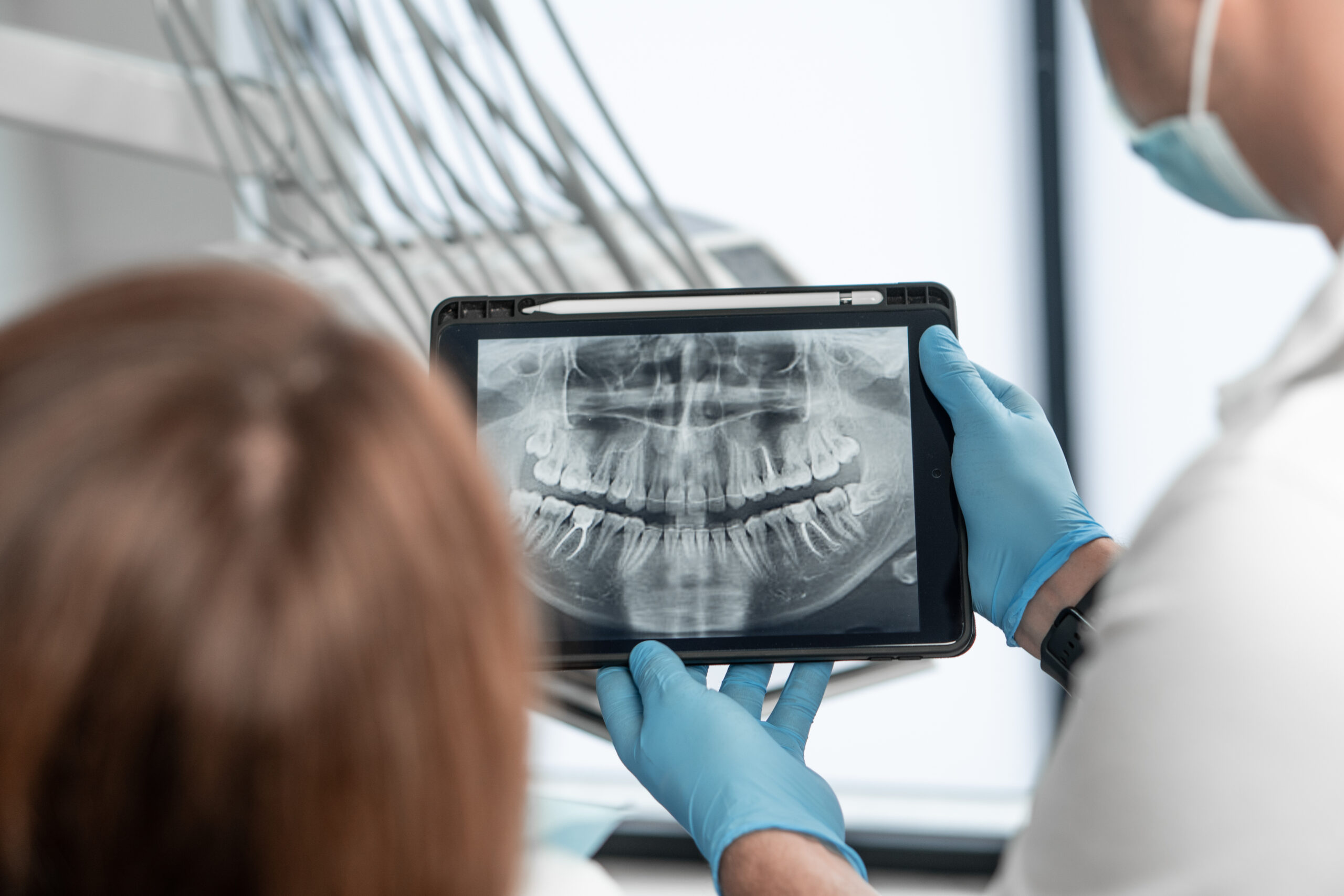
All dental schools teach a system for doing a clinical examination. The goal is typically to gather as much information about current clinical conditions as possible, as efficiently as possible. It is an important aspect of patient care. The science of the exam is useful, but it misses the art of the examination. In my experience, it is often a missed opportunity

In dentistry, we are always trying to figure out the best way to influence our patients to make healthy choices for themselves. When I left Hygiene school, I thought it would be simple to influence patients. I thought that if I did a good examination, a good diagnosis, and then made a good presentation, patients would go ahead and do the treatment.

What I experienced when I was in practice was different. Over time, multiple conversations, and multiple interactions—in their own time and in their own way, patients would move forward with treatment. Sometimes it seemed random, but what I’ve come to understand now is that every interaction was an opportunity to influence the patient.

Every single interaction, with every single patient, by every single member of the dental team is an opportunity to influence.

I think most of us have learned over the years to be skillful at providing information. We know how to “Teach and Tell” what we are finding and recommending. But there is an aspect of that process that has to do with experience. What we have not always paid attention to is how we can go beyond information to create learning experiences for our patients.

When I see a baby touching grass, I imagine that the experience of learning about grass through the senses is entirely different than learning by being told about grass. Creating opportunities for people to interact physically with their own bodies is an opportunity we have in a number of different situations. We can do this during a consultation, but we really have this opportunity during an examination.

If we place priority on effectiveness over efficiency, we will do our exams with the intention of creating physical-sensory experiences, which can be as simple as having them touch their muscles as they touch their teeth together, sliding their jaw forward and side to side, finding a relaxed jaw position, tapping their teeth together, clenching, feeling fremitus with their tongue or finger, feeling the difficulty of flossing between tightly packed teeth, and taking us on a tour of their mouth in a mirror while telling us about their concerns. It’s natural to say, “Tell me more about that. Show me where.”

Consider the new patient exam as the initiation of an experiential learning process to influence our patients to make healthy choices.

The new patient exam is not “the one” opportunity we will have to influence patients. We’ll have many more opportunities, but it sets the tone for every conversation you will have with your patients about their health, about the conditions present in their mouth, about the implications of what is going on in their mouth, and some of the choices they might be able to make.

Job one is to engage the patient in discovering just how intriguing their mouth is and why the health of its various components matter for long-term comfort, function, beauty, and overall health.

Related Course

E1: Aesthetic & Functional Treatment Planning at the Chicago Midwinter Dental Meeting

DATE: February 20 2025 @ 7:00 am - February 22 2025 @ 8:00 pm

Location: Chicago Midwinter Meeting

CE HOURS:

Transform your experience of practicing dentistry, increase predictability, profitability and fulfillment. The Essentials Series is the Key, and Aesthetic and Functional Treatment Planning is where your journey begins.  Following a system of…

Learn More>

About Author

User Image
Mary Osborne RDH

Mary is known internationally as a writer and speaker on patient care and communication. Her writing has been acclaimed in respected print and online publications. She is widely known at dental meetings in the U.S., Canada, and Europe as a knowledgeable and dynamic speaker. Her passion for dentistry inspires individuals and groups to bring the best of themselves to their work, and to fully embrace the difference they make in the lives of those they serve.

FIND A PANKEY DENTIST OR TECHNICIAN

I AM A
I AM INTERESTED IN

VIEW COURSE CALENDAR

Increased Periodontal Disease Risk from Androgen Deprivation Cancer Therapy

June 5, 2023 Lee Ann Brady DMD

I know we are all familiar with the devastating effects cancer therapy can have on our patient’s oral health. We think of chemo and radiation and know this can increase the risk of caries and root resorption. A very common cancer therapy called Androgen Deprivation Therapy can have a significant impact on periodontal risk.

This therapy is an injectable medication given to a significant number of men who have been diagnosed with prostate cancer. It is a commonly chosen therapy because, on the medical side, it is one of the medications with the lowest impact on the patient’s life and ability to more forward with other medical procedures.

Now that it is common among our older male dental patients, I’ve been reading about it. It turns out that this therapy does significantly increase the periodontal risk of bone loss.

When they study males receiving chemotherapy for prostate cancer, 80%+ of the patients on Androgen Deprivation Therapy develop periodontal bone loss. Less than 10% of the males receiving different chemotherapies develop periodontal bone loss.

This is interesting because we think of periodontal disease as being driven by our immune system and that some patients are hyperresponsive to the bacteria in their mouths. In this case, the origin of periodontal risk appears to be different but there is no argument that the risk is there.

Knowing our patients’ medical histories is important yet again! When you do your med-history reviews with your patients, ask them if they are receiving cancer therapy and review the drugs they are on.

As soon as we know a patient is on Androgen Deprivation therapy, we need to start the preventative high-recall, high-maintenance process. Take them to three-month recalls instead of six-month recalls. Apply chlorhexidine varnish and start home hygiene protocols–brushing with baking soda…hydrogen peroxide…all the things we normally advise for our patients when we know they are at high periodontal risk.

Start this as soon as possible even though the patient is presenting with a healthy mouth. The risk percentile is in the 80s, so we can expect to see loss of mucogingival attachment, deepening pocket development, and bone loss.

Related Course

E3: Restorative Integration of Form & Function

DATE: January 12 2025 @ 8:00 am - January 16 2025 @ 2:30 pm

Location: The Pankey Institute

CE HOURS: 41

Dentist Tuition: $ 7400

Single Occupancy with Ensuite Private Bath (per night): $ 345

THIS COURSE IS SOLD OUT Understanding that “form follows function” is critical for knowing how to blend what looks good with what predictably functions well. E3 is the phase of…

Learn More>

About Author

User Image
Lee Ann Brady DMD

Dr. Lee Ann Brady is passionate about dentistry, her family and making a difference. She is a general dentist and owns a practice in Glendale, AZ limited to restorative dentistry. Lee’s passion for dental education began as a CE junkie herself, pursuing lots of advanced continuing education focused on Restorative and Occlusion. In 2005, she became a full time resident faculty member for The Pankey Institute, and was promoted to Clinical Director in 2006. Lee joined Spear Education as Executive VP of Education in the fall of 2008 to teach and coordinate the educational curriculum. In June of 2011, she left Spear Education, founded leeannbrady.com and joined the dental practice she now owns as an associate. Today, she teaches at dental meetings and study clubs both nationally and internationally, continues to write for dental journals and her website, sits on the editorial board of the Journal of Cosmetic Dentistry, Inside Dentistry and DentalTown Magazines and is the Director of Education for The Pankey Institute.

FIND A PANKEY DENTIST OR TECHNICIAN

I AM A
I AM INTERESTED IN

VIEW COURSE CALENDAR

Positioning Peg Laterals & Undersized Lateral Incisors for Optimal Aesthetics 

April 14, 2023 Lee Ann Brady DMD

When restoring peg laterals and laterals that are undersized, great goals are to optimize the final aesthetics and not have to do any tooth preparation prior to adding restorative material. In this blog, I’d like to discuss where we should have the orthodontist optimally position the laterals prior to restoration.

True Peg Laterals

In the case of a true peg lateral, I think of the tooth like I would an implant abutment. In my mind’s eye, I visualize the tooth as a fixture with an abutment on it.

When I talk with the orthodontist, I communicate that I want a minimum of 1 mm and a maximum of 1.5 mm of space between the mesial on the lateral incisor and the distal on the central incisor.

If there is excess space, it is going to be on the distal. We always hide excess space or insufficient space on the distal side of an upper anterior tooth. We always want to perfect the effect on the mesial so we achieve a perfect emergence profile.

And then I communicate that I want the labial of that peg lateral to be positioned about 1 mm to the lingual of where the final facial of the tooth position will be so that I can add material–composite or ceramic, without having to prep the tooth. This position is going to ideally position the free gingival margin of the tooth exactly where I want it based on the free gingival margin of the canine and central incisor. The CEJ is going to be placed exactly where I want the CEJ.

Undersized Lateral Incisors

Often, we have lateral incisors that are not true peg laterals. They’re just undersized lateral incisors. In this case, we must do a thought process about how much restorative material will be added and calibrate how much forward dimension will be added to the tooth. If I’m going to have .5 mm of material on the labial, then I will have the orthodontist position the tooth .5 mm lingually.

If the emergence profile is perfect, then the orthodontist should make it touch the central and all the added material will go on the distal. If not, then a little material will be added to the mesial and to the distal.

Often, for me, the process is thinking, “Where do I want to add restorative material and how much material do I want to add?” Then, I think about where to position the tooth in the space so I will not need to remove any of the tooth structure.

Related Course

E1: Aesthetic & Functional Treatment Planning

DATE: December 11 2025 @ 8:00 am - December 14 2025 @ 2:30 pm

Location: The Pankey Institute

CE HOURS: 39

Dentist Tuition: $ 6800

Single Occupancy with Ensuite Private Bath (Per Night): $ 345

Transform your experience of practicing dentistry, increase predictability, profitability and fulfillment. The Essentials Series is the Key, and Aesthetic and Functional Treatment Planning is where your journey begins.  Following a system of…

Learn More>

About Author

User Image
Lee Ann Brady DMD

Dr. Lee Ann Brady is passionate about dentistry, her family and making a difference. She is a general dentist and owns a practice in Glendale, AZ limited to restorative dentistry. Lee’s passion for dental education began as a CE junkie herself, pursuing lots of advanced continuing education focused on Restorative and Occlusion. In 2005, she became a full time resident faculty member for The Pankey Institute, and was promoted to Clinical Director in 2006. Lee joined Spear Education as Executive VP of Education in the fall of 2008 to teach and coordinate the educational curriculum. In June of 2011, she left Spear Education, founded leeannbrady.com and joined the dental practice she now owns as an associate. Today, she teaches at dental meetings and study clubs both nationally and internationally, continues to write for dental journals and her website, sits on the editorial board of the Journal of Cosmetic Dentistry, Inside Dentistry and DentalTown Magazines and is the Director of Education for The Pankey Institute.

FIND A PANKEY DENTIST OR TECHNICIAN

I AM A
I AM INTERESTED IN

VIEW COURSE CALENDAR

Making Endodontic Diagnosis More Accurate 

February 13, 2023 Lee Ann Brady DMD

It can often be difficult to make an accurate endodontic diagnosis when patients present with tooth pain. We want to be able to get clear results from diagnostic testing so we can feel confident in our treatment recommendations.

One of the things I know to be true for my office and for many of our offices is the challenge of getting clear endodontic diagnostic information. One of the things that can cloud our diagnosis is the effect of over-the-counter medications.

To get accurate endodontic information, the patient must not take pain medication or anti-inflammatory medication in the 8 to 10 hours before you are doing your diagnosis. So, we need to ask patients if they have taken any Tylenol, Advil, or Aleve. We also need to think of patients who are taking nonsteroidal anti-inflammatories on a general basis. These are patients who are not taking them for the tooth but on a routine basis for other reasons such as arthritis.

If I am going to refer the patient to an endodontist and they are going to continue the diagnostic process, I want to coach the patient to not take any pain medication or anti-inflammatory medication for about 8 hours before that appointment. Otherwise, they may not be able to provide the accurate information needed for an accurate diagnosis and most appropriate treatment.

This is something I have passed on to the team members who answer the phone and schedule appointments in my practice. When someone calls to schedule an appointment to diagnose their discomfort, we tell them to do us a favor and not take any more pain medication or anti-inflammatory medication before we see them. Ideally, any of these drugs will be out of the patient’s system before the patient arrives.

Note that your patients who suffer from chronic inflammatory pain conditions such as bursitis, arthritis, and fibromyalgia are often prescribed anti-inflammatory medications that are long-lasting, for example, Celebrex and Meloxicam. These drugs are taken every 24 hours, so their effects last longer and pose a greater risk of clouding a pulpal diagnosis.

Related Course

Direct Composite: Predictable, Easy and Beautiful

DATE: October 25 2024 @ 8:00 am - October 26 2024 @ 4:00 pm

Location: The Pankey Institute

CE HOURS: 14

Regular Tuition: $ 2195

Single Occupancy with Ensuite Private Bath (per night): $ 290

Achieving Predictable and Stunning Anterior Results Direct composite is something we do every day in our practices. Yet, they can sometimes be frustrating when we don’t get a tight contact…

Learn More>

About Author

User Image
Lee Ann Brady DMD

Dr. Lee Ann Brady is passionate about dentistry, her family and making a difference. She is a general dentist and owns a practice in Glendale, AZ limited to restorative dentistry. Lee’s passion for dental education began as a CE junkie herself, pursuing lots of advanced continuing education focused on Restorative and Occlusion. In 2005, she became a full time resident faculty member for The Pankey Institute, and was promoted to Clinical Director in 2006. Lee joined Spear Education as Executive VP of Education in the fall of 2008 to teach and coordinate the educational curriculum. In June of 2011, she left Spear Education, founded leeannbrady.com and joined the dental practice she now owns as an associate. Today, she teaches at dental meetings and study clubs both nationally and internationally, continues to write for dental journals and her website, sits on the editorial board of the Journal of Cosmetic Dentistry, Inside Dentistry and DentalTown Magazines and is the Director of Education for The Pankey Institute.

FIND A PANKEY DENTIST OR TECHNICIAN

I AM A
I AM INTERESTED IN

VIEW COURSE CALENDAR

Why Do Prepared Teeth Discolor?

December 23, 2022 Lee Ann Brady DMD

If the color of a prepared tooth darkens between the time you prepare it and the restoration is seated, the aesthetics of the final restoration can be impaired. This frustrating situation can be eliminated by knowing the causes of discoloration and what to do when planning treatment and prepping the tooth.

There are two processes that cause prepared teeth to discolor to a darker shade:
  1. Pulpal necrosis
  2. Chemical interaction between liquid vasoconstrictors and bacteria in the dentin tubules

Note that both processes can continue to further darken dentin weeks to months after you have seated the restoration. For more predictable aesthetic results, I learned some time ago to do the following.

Assess pulpal vitality first.

I am highly cautious when planning significant restorative treatment such as crown and bridge. Before prepping teeth, I review CBCT radiographs to make sure there are no pulpal health issues that need to be treated first. Like most dentists, I do not have CBCT imaging in my own practice, but I do have access to CBCT imaging via a collaborative relationship with a nearby specialist.

Use retraction paste instead of liquid vasoconstrictors for hemostasis.

Because the chemistry in liquid-viscosity vasoconstrictors can interact with bacteria in the dentin tubules to darken the dentin, I use retraction paste when I need hemostasis.

For me, these two seemingly simple steps are important ones when seeking optimal aesthetic results.

Related Course

TMD & Orofacial Pain: Managing Complex Patients

DATE: January 29 2025 @ 8:00 am - February 2 2025 @ 1:00 pm

Location: The Pankey Institute

CE HOURS: 37

Dentist Tuition: $ 7200

Single Occupancy with Ensuite Private Bath (per night): $ 345

TMD patients present with a wide range of concerns and symptoms from tension headaches and muscle challenges to significant joint inflammation and breakdown. Accurate thorough diagnosis is the first step…

Learn More>

About Author

User Image
Lee Ann Brady DMD

Dr. Lee Ann Brady is passionate about dentistry, her family and making a difference. She is a general dentist and owns a practice in Glendale, AZ limited to restorative dentistry. Lee’s passion for dental education began as a CE junkie herself, pursuing lots of advanced continuing education focused on Restorative and Occlusion. In 2005, she became a full time resident faculty member for The Pankey Institute, and was promoted to Clinical Director in 2006. Lee joined Spear Education as Executive VP of Education in the fall of 2008 to teach and coordinate the educational curriculum. In June of 2011, she left Spear Education, founded leeannbrady.com and joined the dental practice she now owns as an associate. Today, she teaches at dental meetings and study clubs both nationally and internationally, continues to write for dental journals and her website, sits on the editorial board of the Journal of Cosmetic Dentistry, Inside Dentistry and DentalTown Magazines and is the Director of Education for The Pankey Institute.

FIND A PANKEY DENTIST OR TECHNICIAN

I AM A
I AM INTERESTED IN

VIEW COURSE CALENDAR

Is It Time to Review Your Treatment Protocol for Traumatized Teeth?

December 12, 2022 Lee Ann Brady DMD

About a year ago, in my office, we reviewed our protocol for managing and treating patients with traumatized teeth. We reviewed the literature to learn how we could improve how we help our adult patients who have avulsed or mobile teeth following a traumatic event.

Antibiotics? Yes

One of the new things we read and thought about was whether to put the patient on antibiotics. We added this to our protocol—the patient goes on antibiotics for seven days after learning what antibiotics the patient can take, i.e., will not likely cause an allergic reaction.

Splinting? Yes

The literature now recommends splinting the traumatized teeth for two weeks and then removing the split after two weeks. Although there has been a conversation over the years about whether to splint or not to splint and if splinting has anything to do with the teeth ankylosing or resorption, the current recommendation is to splint but for just two weeks.

Improved Counseling of Our Patients? Yes

We learned that three common sense items needed to be reviewed with our patients, because it is easy to retraumatize teeth, and patients easily forget to be attentive to personal “gentleness.”

  • We added to our protocol list counseling the patient to go on a soft diet that does not require biting down for three to four days, then longer if they sense the tooth roots are still mobile.
  • Similarly, we added counseling the patient to do gentle mouth cleaning. They should brush traumatized teeth very, very gently so as not to re-traumatize or move them.
  • We also added to our list making sure patients understand the importance of follow-up visits and radiography to track the health of the traumatized teeth.

Following the Health of the Teeth

We use periapical or CBT radiography to follow the teeth at one month after the initial trauma and again after two months, four months, and six months. If there appears to be healthy pulp and attachment of the teeth to the bone and connective tissue at six months, we can extend the time between making new images.

Related Course

Surgically Facilitated Orthodontic Therapy: An Interdisciplinary Approach

DATE: April 26 2024 @ 2:00 pm - April 26 2024 @ 4:00 pm

Location: Online

CE HOURS: 2

Surgically Facilitated Orthodontic Therapy: An Interdisciplinary Approach Patients seeking ideal esthetics may require a more sophisticated diagnosis and treatment plan than what restorative dentistry or conventional orthodontic treatment may offer…

Learn More>

About Author

User Image
Lee Ann Brady DMD

Dr. Lee Ann Brady is passionate about dentistry, her family and making a difference. She is a general dentist and owns a practice in Glendale, AZ limited to restorative dentistry. Lee’s passion for dental education began as a CE junkie herself, pursuing lots of advanced continuing education focused on Restorative and Occlusion. In 2005, she became a full time resident faculty member for The Pankey Institute, and was promoted to Clinical Director in 2006. Lee joined Spear Education as Executive VP of Education in the fall of 2008 to teach and coordinate the educational curriculum. In June of 2011, she left Spear Education, founded leeannbrady.com and joined the dental practice she now owns as an associate. Today, she teaches at dental meetings and study clubs both nationally and internationally, continues to write for dental journals and her website, sits on the editorial board of the Journal of Cosmetic Dentistry, Inside Dentistry and DentalTown Magazines and is the Director of Education for The Pankey Institute.

FIND A PANKEY DENTIST OR TECHNICIAN

I AM A
I AM INTERESTED IN

VIEW COURSE CALENDAR

Not Every Endodontically Treated Tooth Needs a Crown

August 22, 2022 Lee Ann Brady DMD

Not every tooth that has been endodontically treated requires a crown to insure it has great longevity and doesn’t crack or fracture.

Molars

Very clearly the literature supports that molar teeth in the posterior absolutely must have four cusp coverage—a four-cusp onlay or a full coverage crown. We are trying to use the phenomenon of containment with strong ceramic or metal material around the entire circumference of the tooth. We are holding the buccal and lingual together and replacing the top of the root chamber, so the tooth doesn’t fracture.

Bicuspids

Bicuspids that have been endodontically treated do not need two-cusp coverage if there have been no previous restorations and the endo access is very conservative. In the case of a premolar that has never had an MO, a DO, or an MOD, and has a tiny access hole, you can do a composite buildup or chamber retaining composite restoration. If the patient has high functional risk, a reasonable decision would be to restore the tooth with an onlay or crown

Anterior Teeth

There is no scientific support for doing a crown on an anterior tooth just because it has had endodontic therapy. We do a crown on an anterior tooth that has had endo when it is already structurally compromised, for example with previous mesial lingual and distal lingual composite fillings, missing tooth structure, and significant structural compromise between the endo access and other restorations.

Related Course

E4: Posterior Reconstruction and Completing the Comprehensive Treatment Sequence

DATE: May 2 2024 @ 8:00 am - May 6 2024 @ 2:30 pm

Location: The Pankey Institute

CE HOURS: 44

Dentist Tuition : $ 7300

night with private bath: $ 290

This Course Is Sold Out! The purpose of this course is to help you develop mastery with complex cases involving advanced restorative procedures, precise sequencing and interdisciplinary coordination. Building on…

Learn More>

About Author

User Image
Lee Ann Brady DMD

Dr. Lee Ann Brady is passionate about dentistry, her family and making a difference. She is a general dentist and owns a practice in Glendale, AZ limited to restorative dentistry. Lee’s passion for dental education began as a CE junkie herself, pursuing lots of advanced continuing education focused on Restorative and Occlusion. In 2005, she became a full time resident faculty member for The Pankey Institute, and was promoted to Clinical Director in 2006. Lee joined Spear Education as Executive VP of Education in the fall of 2008 to teach and coordinate the educational curriculum. In June of 2011, she left Spear Education, founded leeannbrady.com and joined the dental practice she now owns as an associate. Today, she teaches at dental meetings and study clubs both nationally and internationally, continues to write for dental journals and her website, sits on the editorial board of the Journal of Cosmetic Dentistry, Inside Dentistry and DentalTown Magazines and is the Director of Education for The Pankey Institute.

FIND A PANKEY DENTIST OR TECHNICIAN

I AM A
I AM INTERESTED IN

VIEW COURSE CALENDAR

Functional Risk Part 3 – Occlusal Therapy 

July 1, 2022 Lee Ann Brady DMD

Why Occlusal Appliance Therapy Is My First Step Prior to Ortho, Equilibration, or Restorative

Occlusal changes on an appliance are easy and reversible. An appliance can immediately reduce elevator muscle activity and give the patient relief. The patient also experiences what changes to their tooth contacts could provide for them long term. We can test the changes that would be made by ortho, equilibration, and/or restorative.

As reviewed in Part 2 of this series, our goals are to stabilize the joint anatomy and reduce the activity of the elevator muscles because those muscles are what overload the joints and teeth. We also want to slow down the rate of damage to the dentition and move that rate back to a more age-appropriate pace. We also may need to reorganize a patient’s occlusion to manage occlusal forces to ensure restorations that last.

Removing Posterior Contacts Does Not Work for Every Patient

Over my years of clinical practice, I have found that changing the occlusion does reduce functional risk for most patients. But we all have patients with perfect occlusion who present with TMD symptoms. We have some patients who continue to parafunction after we move them into immediate posterior disclusion.

Studies show that proprioception causes the elevator muscles to engage in only 80 to 85% of the population. This means that when the brain receives the signal that teeth are touching, the brain elevates the masseter muscles in 80 to 85% of people. Tooth contact is the trigger. Because this proprioception does not occur for 15 to 20% of the population, it is not the universal trigger for excessive loading.

Over my years in clinical practice, I have learned there is nothing I can do that is 100% dependable to stop a patient from para-functioning. Some of my patients continue to excessively load after posterior contacts are removed. Their functional risk does not diminish.

If we cannot reduce elevator force and redistribute force enough on an occlusal appliance to eliminate or at least relieve TMD symptoms, then occlusal therapy via ortho, equilibration, or restorative will not satisfactorily help the patient. We will need to turn to other forms of therapy.

Other modalities I use are BOTOX to deactivate muscles, massage therapy, and physical therapy. There are also systemic medications, cold lasers, and TENS therapy we can use to reduce the activity of the muscles or reduce inflammation in the muscles and joints. Sometimes one modality will alleviate symptoms for a while and when symptoms return, we can try it again or try another modality.

An Exercise to Identify the Patients Who May Not Benefit from Occlusal Therapy

You can do what I call a poor man’s EMG on yourself by placing your hands on your masseter muscles. Put your back teeth together, clench and release, clench and release, clench and release to see how much masseter activity you have. Then move your teeth into protrusive edge to edge and try to clench a little bit, making sure your back teeth do not touch. If you now have a posterior tooth touching in the edge-to-edge position, then put a pencil or pen between your front teeth to separate your back teeth.

With no back teeth touching and contact on the centrals, try to clench and release two or three times while feeling your masseters. Most of you will find your masseters do not move or move a lot less when no back teeth are touching. Some of you, even with your back teeth separated, can still clench in protrusive and can still increase the muscle activity almost the same amount as when your back teeth touch.

I do this exercise with my patients, but when they move into protrusive, I put a bite stop over their front teeth or have them bite on a Lucia jig we have lined for their bite registration. If you do this test with your patients, you can use an EMG or feel the muscle activity with your hands.

If the patient can still generate almost the same force or the same force with their back teeth separated, you have identified one of the around 15% of people who might not benefit significantly from occlusal therapy. You’ve also identified someone who might not do well on an anterior-only appliance because, if they can generate that same force on just two teeth, they are at risk for those teeth becoming sore and moving.

Interested in Learning More?

The Pankey Institute Essentials courses and multiple focus courses include hands-on exercises and over-the-shoulder training designed to help dentists develop mastery in reducing functional risk and treating TMD symptoms.

Related Course

E1: Aesthetic & Functional Treatment Planning

DATE: October 16 2025 @ 8:00 am - October 19 2025 @ 2:30 pm

Location: Ciao Restaurant

CE HOURS: 39

Dentist Tuition: $ 6800

Single Occupancy with Ensuite Private Bath (Per Night): $ 345

Transform your experience of practicing dentistry, increase predictability, profitability and fulfillment. The Essentials Series is the Key, and Aesthetic and Functional Treatment Planning is where your journey begins.  Following a system of…

Learn More>

About Author

User Image
Lee Ann Brady DMD

Dr. Lee Ann Brady is passionate about dentistry, her family and making a difference. She is a general dentist and owns a practice in Glendale, AZ limited to restorative dentistry. Lee’s passion for dental education began as a CE junkie herself, pursuing lots of advanced continuing education focused on Restorative and Occlusion. In 2005, she became a full time resident faculty member for The Pankey Institute, and was promoted to Clinical Director in 2006. Lee joined Spear Education as Executive VP of Education in the fall of 2008 to teach and coordinate the educational curriculum. In June of 2011, she left Spear Education, founded leeannbrady.com and joined the dental practice she now owns as an associate. Today, she teaches at dental meetings and study clubs both nationally and internationally, continues to write for dental journals and her website, sits on the editorial board of the Journal of Cosmetic Dentistry, Inside Dentistry and DentalTown Magazines and is the Director of Education for The Pankey Institute.

FIND A PANKEY DENTIST OR TECHNICIAN

I AM A
I AM INTERESTED IN

VIEW COURSE CALENDAR

Functional Risk Part 2 – Why Does Changing Occlusion Work?

June 24, 2022 Lee Ann Brady DMD

High functional risk is evidenced in damage to the jaw joints, muscles, or teeth that exceeds what is normal for the person’s age. High functional risk occurs when there has been excessive loading in a compressed period due to what I described in Part 1 of this blog series as macro trauma from an impact event or micro trauma from chronic parafunction.

To manage functional risk and slow down the attrition we are observing, dentists adjust the patient’s occlusion to reduce that load. What are our goals in changing the occlusion and why does changing occlusion work well in most cases?

Functional Risk Management Goals

Whether we are managing functional risk with a bite splint, orthodontics, occlusal adjustment and/or restorative dentistry, we want to:

  • Stabilize joints
  • Stabilize muscles
  • Stabilize dentition
  • Ensure predictable restorative outcomes

We want to stabilize the joint anatomy, the structures of the temporomandibular joints, and reduce the activity of the elevator muscles because they are what overloads the joints and teeth and can be used with so much force that the muscles injure themselves. We also want to slow down the rate of damage to the dentition and move that rate back to a more age-appropriate pace. Often, we need to reorganize a patient’s occlusion to manage occlusal forces to ensure predictable restorative outcomes that last.

Why Changing Occlusion Works

Dr. Bob Barkley said, “Our job as dentists is to help our patients get worse at the slowest possible rate.” And that is what occlusal therapy does. When we change the occlusion, we are minimizing the force applied across the tops of the teeth and redistributing the force applied across the tops of the teeth.

The Science Behind This

In the dental literature, most studies are based on electromyographic activity. The patient is given a false occlusion on an appliance and the occlusion is altered. EMG activity is read when the teeth touch under many variations. By adding a premolar contact, the activity of the elevator muscles doubles. When a second molar contact is added, the activity of the elevator muscles rises five to ten times. A key article is Influence of variations in anteroposterior occlusal contacts on electromyographic activity by Arturo Manns, et al.

The EMG studies have demonstrated posterior tooth contacts produce the greatest load, so to minimize load, we eliminate posterior tooth contact. For most patients, we can’t get them to immediately disclude on their centrals, but we can get them to disclude on the canines, and we want to pass that off as fast as we can to the centrals. The term canine guidance or anterior guidance refers to the absence of posterior contacts. Instead of saying canine guidance or anterior guidance, many people use the term immediate posterior disclusion.

Note: The two-to-ten-times-more force recorded on an EMG study appliance is likely much lower than the force applied by muscles adapted to para-functioning over years and years. Those adapted muscles have become larger, more fibrous, and can generate more force.

Removing Posterior Contacts Does Not Work for Every Patient

Over my years of clinical practice, I have found that changing the occlusion does reduce functional risk for most patients. But we all have patients with perfect occlusion who present with TMD symptoms. We have some patients who continue to parafunction after we move them into immediate posterior disclusion.

Studies show that proprioception causes the elevator muscles to engage in only 80 to 85% of the population. This means that when the brain receives the signal that teeth are touching, the brain elevates the masseter muscles in 80 to 85% of people. Tooth contact is the trigger. Because this proprioception does not occur for 15 to 20% of the population, it is not the universal trigger for excessive loading.

Over my years in clinical practice, I have learned there is nothing I can do that is 100% dependable to stop a patient from para-functioning. Some of my patients continue to excessively load after posterior contacts are removed. This will be revisited in Part 3 – Management Modalities.

Related Course

E1: Aesthetic & Functional Treatment Planning

DATE: June 20 2024 @ 8:00 am - June 23 2024 @ 2:30 pm

Location: The Pankey Institute

CE HOURS: 39

Dentist Tuition: $ 6500

Single Occupancy Room with Ensuite Bath (Per Night): $ 290

Transform your experience of practicing dentistry, increase predictability, profitability and fulfillment. The Essentials Series is the Key, and Aesthetic and Functional Treatment Planning is where your journey begins.  Following a system of…

Learn More>

About Author

User Image
Lee Ann Brady DMD

Dr. Lee Ann Brady is passionate about dentistry, her family and making a difference. She is a general dentist and owns a practice in Glendale, AZ limited to restorative dentistry. Lee’s passion for dental education began as a CE junkie herself, pursuing lots of advanced continuing education focused on Restorative and Occlusion. In 2005, she became a full time resident faculty member for The Pankey Institute, and was promoted to Clinical Director in 2006. Lee joined Spear Education as Executive VP of Education in the fall of 2008 to teach and coordinate the educational curriculum. In June of 2011, she left Spear Education, founded leeannbrady.com and joined the dental practice she now owns as an associate. Today, she teaches at dental meetings and study clubs both nationally and internationally, continues to write for dental journals and her website, sits on the editorial board of the Journal of Cosmetic Dentistry, Inside Dentistry and DentalTown Magazines and is the Director of Education for The Pankey Institute.

FIND A PANKEY DENTIST OR TECHNICIAN

I AM A
I AM INTERESTED IN

VIEW COURSE CALENDAR

Functional Risk Part 1 – What Causes It?

June 20, 2022 Lee Ann Brady DMD

I like to think that I have three things I provide to my dental patients:

  1. Risk assessment – helping them understand and fully own risk factors for their long-term dental health
  2. Risk management – helping them understand what they could do to manage that risk
  3. Damage repair – definitively treating a risk that was not completely managed

Many dentists do not pay attention to occlusion unless it is a problem for the patient or unless it becomes an issue in treating the patient. As I tell dentists in Essentials 1: Aesthetic& Functional Treatment Planning, assessing functional risk is as important to me as assessing other risks, such as caries or periodontal disease. I want to find the signs of functional risk, so if a patient has higher risk of damaging teeth from excessive loading, I can help the patient understand that risk and the options for managing it.

Functional Risk Assessment

In a previous blog, Occlusal Wear Part 1: Is it advancing? How fast?, I shared the mental game I play with every patient and the ways in which I document wear changes. With every patient, I ask myself, “Is the wear I see on the teeth normal for the patient’s age? Is it advancing at a pathological rate?”

I categorize patients in one of three functional risk categories:

  • Mild
  • Moderate
  • High

The patients I place in the high-risk category are those whose functional wear and tear is more than it should be for their age. Their teeth are breaking down noticeably faster than the average rate.

In my practice, we measure from the CEJ to the incisal edge of several teeth with wear. We take the measurement on the mid-facial and record it on the patient’s perio chart. At subsequent appointments, we can now repeat these measurements and have clear data showing that the process is continuing. Other great ways to document tooth wear are with photography and digital impressions. We compare scans months later and get a precise measurement of the change.

What causes someone to be at higher functional risk?

A lot of our patients have true TMD. What causes them to become symptomatic–where they have muscle issues, limited range of motion, jaw fatigue or joints trauma, myofascial pain, and they are breaking down their teeth? There are two primary causes: macro trauma and micro trauma.

  1. Macro trauma can cause a temporary injury to the temporomandibular system that then sets up chronic problems in the joints and muscles. This could be due to a car accident or sports incident. I have a macro-trauma patient who was hit was a lacrosse stick, another that was elbowed in the jaw during a basketball game, and a cheerleader who fell off a human tower.
  2. Micro trauma is what dentists call parafunction. This occurs when people put their teeth together outside the normal ways teeth touch when eating, speaking, and swallowing. We think of clenching (both static clenching and power wiggling), grinding, and tapping teeth together. We think of patients who bite their fingernails or chew on the inside of their cheeks or lips. There are lots of types of parafunctional activities. The force generated by the elevator muscles and how much of the time the muscles are overloaded leads to muscle symptoms. Accumulative force causes the excessive wear we see on teeth and damage to jaw joints.

To dentists, I say:

There are many people who have textbook malocclusions, and yet they have healthy teeth and joints. They don’t touch their teeth together outside of eating, speaking, and swallowing. There are many people with perfect occlusions who have TMD symptoms. Malocclusions don’t cause functional risk. Malocclusions don’t cause TMD. The essence of the problem is not how the teeth touch but how much they touch.

To patients with micro trauma, I say:

“You are tougher on your teeth than most of my other patients.” Staying away from psychologically negative words like clenching, grinding, and parafunction, I give them the word tougher. And I say, “You are missing more tooth structure than most people of your age.”

It is helpful for them to have this explanation before I recommend risk management strategies and pre-emptive restoration of teeth before they break.

An analogy I use with patients is the human knee. Knees don’t commonly wear out until someone is 60 to 70 years old, but long-distance runners can wear them out much earlier in life with the repetitive force of running. Our patients with parafunction put a lifetime’s worth of wear and tear on their teeth and their muscles and jaw joints in a compressed amount of time. Like a long-distance runner, their masticatory system suffers micro trauma.

It’s helpful to give patients words and analogies (like knees and car engines that wear out due to faster than normal wear and tear). I’ve had patients say to me, “I don’t like having to replace this crown, but as you said, I am tougher on my teeth than most people.”

Related Course

E1: Aesthetic & Functional Treatment Planning

DATE: December 11 2025 @ 8:00 am - December 14 2025 @ 2:30 pm

Location: The Pankey Institute

CE HOURS: 39

Dentist Tuition: $ 6800

Single Occupancy with Ensuite Private Bath (Per Night): $ 345

Transform your experience of practicing dentistry, increase predictability, profitability and fulfillment. The Essentials Series is the Key, and Aesthetic and Functional Treatment Planning is where your journey begins.  Following a system of…

Learn More>

About Author

User Image
Lee Ann Brady DMD

Dr. Lee Ann Brady is passionate about dentistry, her family and making a difference. She is a general dentist and owns a practice in Glendale, AZ limited to restorative dentistry. Lee’s passion for dental education began as a CE junkie herself, pursuing lots of advanced continuing education focused on Restorative and Occlusion. In 2005, she became a full time resident faculty member for The Pankey Institute, and was promoted to Clinical Director in 2006. Lee joined Spear Education as Executive VP of Education in the fall of 2008 to teach and coordinate the educational curriculum. In June of 2011, she left Spear Education, founded leeannbrady.com and joined the dental practice she now owns as an associate. Today, she teaches at dental meetings and study clubs both nationally and internationally, continues to write for dental journals and her website, sits on the editorial board of the Journal of Cosmetic Dentistry, Inside Dentistry and DentalTown Magazines and is the Director of Education for The Pankey Institute.

FIND A PANKEY DENTIST OR TECHNICIAN

I AM A
I AM INTERESTED IN

VIEW COURSE CALENDAR